© PMC | AI
Radiological Subspecialties
Pediatric Radiology
As the name suggests, pediatric radiology is the branch of radiology focused on diagnostic imaging in infants, children, and adolescents. Because a child's body is still growing and developing, both the clinical presentations and the imaging requirements differ significantly from those in adults. A central concern is radiation protection. Not only are children more sensitive to radiation, but they also have a longer life ahead of them during which potential late effects could emerge. For this reason, radiation-free methods such as ultrasound and magnetic resonance imaging are preferred, while X-ray and computed tomography are used only when clearly indicated and at the lowest possible dose. Pediatric radiology is applied across a wide range of situations, including congenital malformations, developmental disorders, infections, injuries, and unexplained symptoms such as abdominal pain or fever without an obvious cause. Imaging must not only be technically adapted to younger patients but must also account for the unique anatomy and dynamics of the growing body. For example, changes in bone growth, maturation processes, and age-dependent normal variants can all be evaluated.
Musculoskeletal Radiology (MSK Radiology)
MSK radiology focuses on imaging of the musculoskeletal system, including bones, joints, muscles, tendons, and ligaments. It is used primarily for complaints such as pain and restricted movement, as well as following injuries, and plays a central role in orthopedics, sports medicine, and trauma surgery. A range of imaging methods is employed to enable precise diagnosis. X-rays generally serve as the first diagnostic step, providing a quick overview of bone structures, possible fractures, and signs of wear. For the assessment of soft tissues, magnetic resonance imaging is particularly well suited, as it depicts ligaments, muscles, tendons, and menisci in detail — making it indispensable in the evaluation of sports injuries. Ultrasound can also be used to assess superficial structures and to observe them in motion.
For complex joint problems, MRI can be extended with MR arthrography, in which a contrast agent is introduced directly into the joint to visualize cartilage, the joint labrum, and fine ligamentous structures in greater detail. Beyond pure diagnostics, musculoskeletal radiology also encompasses minimally invasive, image-guided procedures. These include interventional pain therapy of the spine — such as periradicular therapy at irritated nerve roots and infiltration of the small facet joints — as well as image-guided punctures and infiltrations at other joints. Musculoskeletal imaging is therefore used for both acute injuries, such as cruciate ligament tears, meniscal damage, or muscle injuries, and chronic conditions such as osteoarthritis or persistent back pain.
Thoracic Radiology
Thoracic radiology focuses on imaging of the chest, particularly the lungs, airways, ribs, chest wall, and, in part, the heart. It is used primarily for complaints such as cough, shortness of breath, chest pain, or unexplained infections. The classic chest X-ray is often the first examination performed, as it is quickly available and can provide important clues about common conditions such as pneumonia, fluid accumulation, or structural changes. Computed tomography is frequently used for more precise diagnosis, as it enables a considerably more detailed view of lung structure. When there is suspicion of lung tumors, pulmonary embolism, or complex inflammatory processes, CT provides crucial information, revealing even the smallest changes that may not yet be visible on an X-ray. The scope of thoracic radiological diagnostics is broad: it can determine whether an acute infection is present, whether chronic lung diseases exist, whether tumors are identified, or whether vascular conditions such as pulmonary embolism are present.
Abdominal Radiology
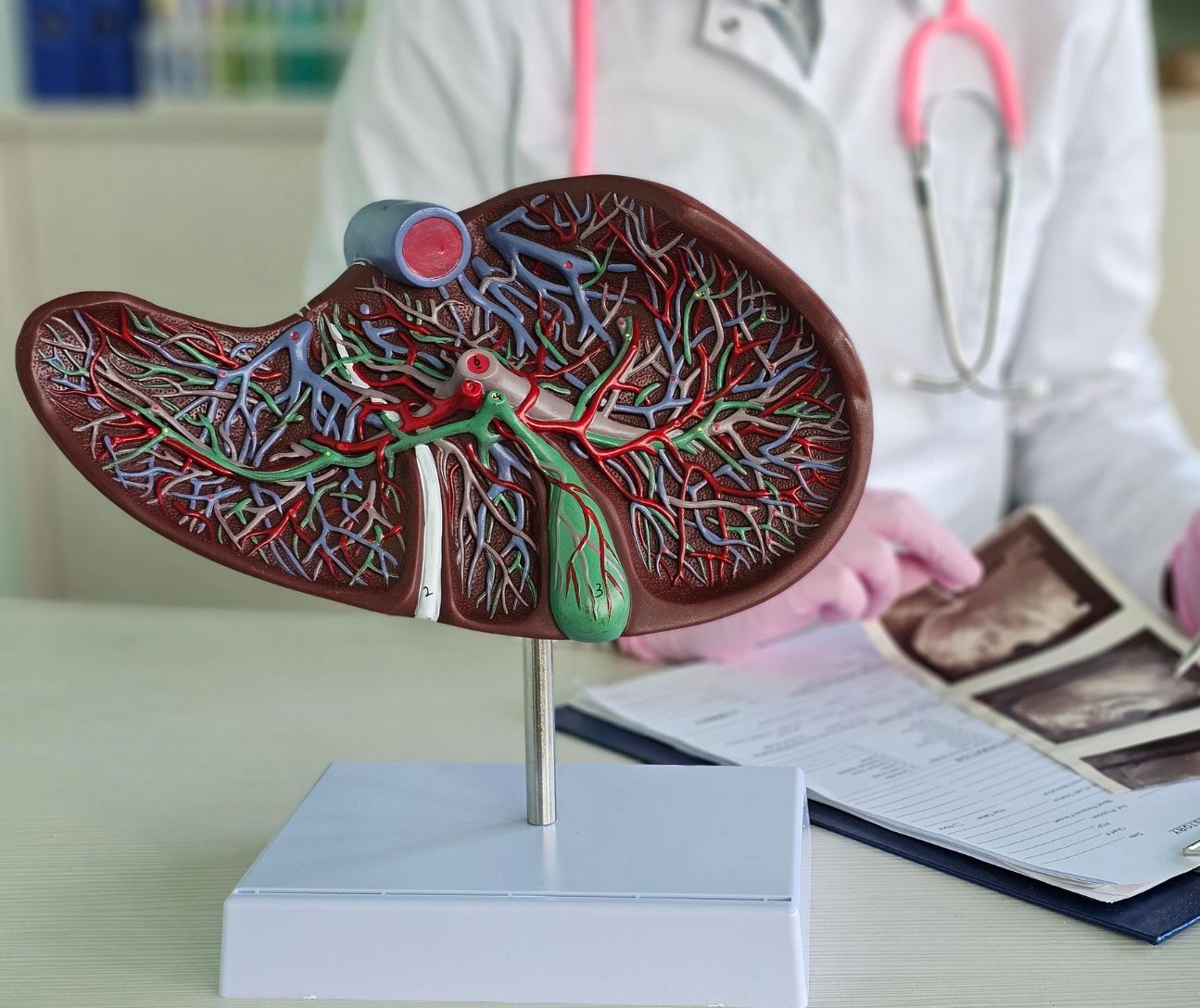
Abdominal radiology encompasses imaging of the abdominal organs, particularly the liver, gallbladder, pancreas, spleen, intestines, kidneys, and urinary bladder. It is used primarily for symptoms such as abdominal pain, digestive disorders, unexplained inflammation, or suspected tumor disease. Ultrasound is often the first imaging method employed, as it is fast, involves no radiation exposure, and is readily available. It enables an initial assessment of organ structure, blood flow, and possible abnormalities such as stones, cysts, or fluid accumulations. For more detailed evaluation, the treating physician may choose computed tomography or magnetic resonance imaging. CT is particularly well suited for urgent clinical questions, such as suspected internal bleeding, inflammation, or injuries. MRI, on the other hand, offers highly detailed visualization of soft tissues and is frequently used for liver disease, tumor diagnostics, or complex clinical questions. Abdominal imaging makes it possible to identify both structural changes and functional correlations — for example, tumors can be localized and assessed in terms of their extent, inflammations can be identified, and circulatory disorders can be detected.
Cardiovascular Radiology
Cardiovascular radiology focuses on imaging of the heart and blood vessels. Computed tomography and magnetic resonance imaging are the primary methods used, supplemented by specialized techniques for vascular visualization. These examinations are employed when cardiovascular disease is suspected — for example, in cases of chest pain, suspected coronary artery disease, vascular narrowing, or congenital heart defects. CT angiography enables rapid, high-resolution visualization of the coronary arteries and can reveal whether narrowing or calcification is present. It is frequently used to assess the risk of heart attacks or to further investigate unexplained symptoms. Cardiac magnetic resonance imaging, in turn, provides detailed information about heart muscle function, blood supply, scarring, and inflammatory changes. The findings obtained allow important conclusions to be drawn about the heart's performance and the condition of the vessels — for example, whether circulatory disorders exist or whether the heart muscle has been damaged. This information is crucial for selecting the appropriate therapy, whether medication, interventional procedures, or surgical measures, and plays a central role in the prevention and treatment of cardiovascular disease.
Experts for this Treatment Method
All Experts in this Department
Show All
- Radiology and radiation therapy
Prof. Dr. Hendrik Wolff
Life Beam
- Radiology and radiation therapy
Prof. Dr. Marc Regier
Radiologie München im Alten Hof
- Radiology and radiation therapy
Prof. Dr. Christopher Herzog
Radiologie im Rotkreuzklinikum
- Radiology and radiation therapy