© Magnific
Diagnostic Radiology
X-Ray Diagnostics
Conventional X-ray imaging is typically the first and most readily available diagnostic tool. It works by directing X-rays through the body, where they are absorbed to varying degrees depending on the type of tissue. Bones appear noticeably brighter than soft tissue because they absorb more radiation. X-rays are used primarily when a physician needs an initial overview — for example, when a bone fracture, joint injury, degenerative changes, misalignment, or lung disease is suspected. A chest X-ray is also commonly taken in cases of persistent cough, shortness of breath, or suspected pneumonia, and X-rays remain a reliable diagnostic tool in dentistry as well. The main advantages of X-ray imaging lie in its speed, wide availability, and relatively straightforward execution. However, its diagnostic value is limited when it comes to fine soft tissue structures, ligaments, nerves, or early inflammatory changes — situations in which computed tomography (CT) or magnetic resonance imaging (MRI) is often needed for a more precise assessment.
Computed Tomography (CT)
Computed tomography is also an X-ray-based procedure, but rather than producing simple projection images, it generates high-resolution cross-sectional images of the body. This allows organs, bones, vessels, and internal injuries to be depicted far more precisely — which is especially important in emergency medicine, for example in cases of polytrauma, suspected internal bleeding, stroke, pulmonary embolism, or complex bone fractures. CT also plays a key role in oncological conditions, abdominal inflammation, and inconclusive findings. A treating physician will turn to CT whenever a highly accurate picture of the overall situation is required. Unlike conventional X-rays, CT displays structures layer by layer, enabling a significantly better spatial assessment. This makes it possible to determine, for instance, whether bleeding is present, whether an organ has been partially injured, whether a tumor has spread, or whether a vascular narrowing or occlusion exists. The conclusions drawn from a CT scan are therefore often very concrete and decisive for the next steps — whether that means drug therapy, close monitoring, or surgery.
Magnetic Resonance Imaging (MRI)
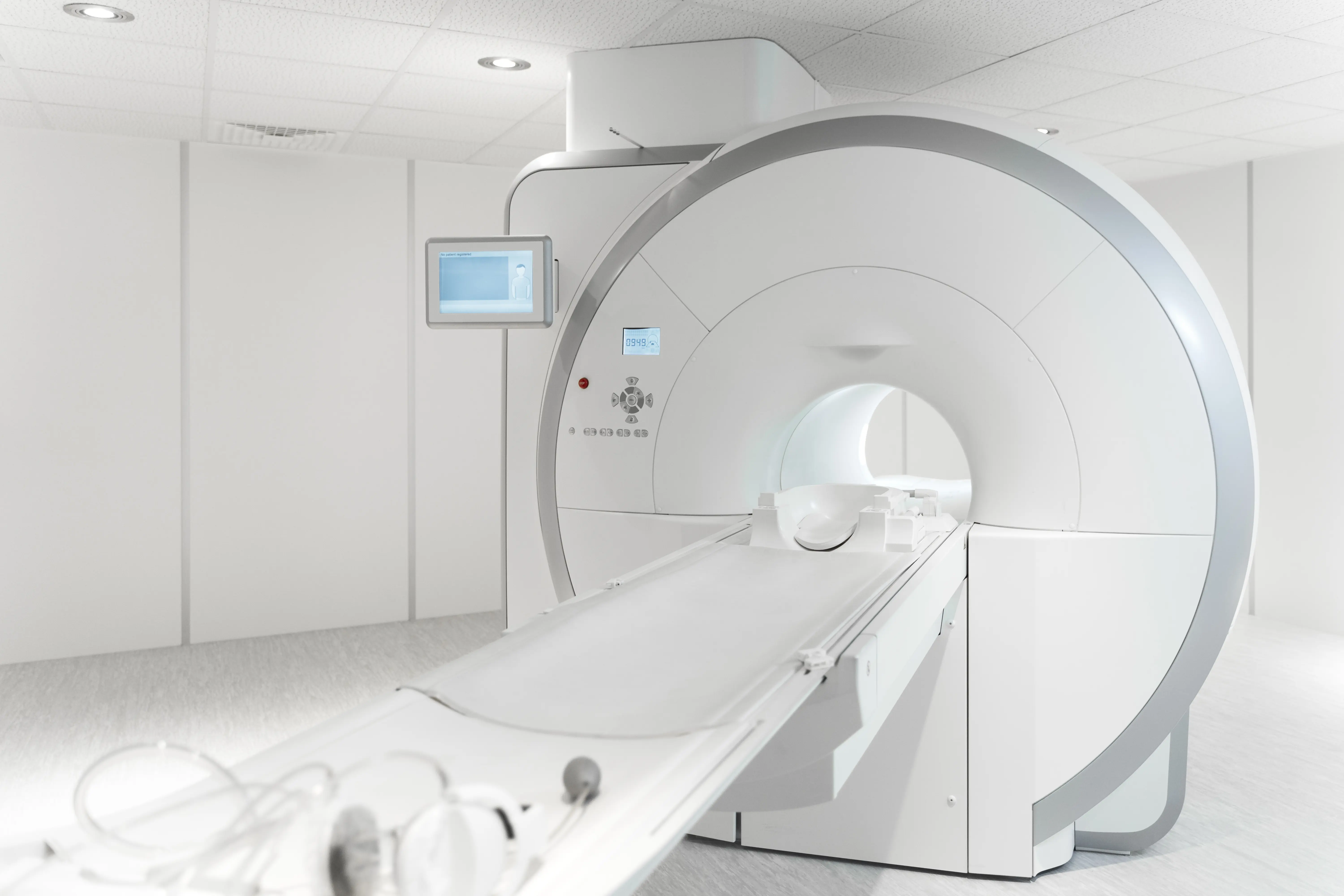
Magnetic resonance imaging does not use X-rays; instead, it relies on strong magnetic fields and radio waves. This technique is particularly well suited for depicting soft tissues in great detail, which is why MRI is used primarily for conditions affecting the brain, spinal cord, intervertebral discs, joints, muscles, tendons, and ligaments, as well as in tumor diagnostics. MRI is also the preferred choice when symptoms cannot be adequately explained by an X-ray — for example, in cases of persistent back pain with suspected disc herniation, or knee injuries with possible damage to the meniscus or cruciate ligaments. Neurological symptoms such as paralysis, sensory disturbances, or unexplained headaches may equally prompt a physician to order an MRI. The key advantage of MRI lies in its exceptional soft tissue resolution. This not only reveals whether a change is present, but also which tissue is affected and how severe the damage is. It can show, for instance, whether a ligament is partially or completely torn, whether a disc is pressing on a nerve, or whether a tumor has grown into surrounding tissue. Bone marrow changes that would be invisible on a conventional X-ray can also be detected with MRI. In orthopedics, neurology, and oncology in particular, MRI findings play an important role in treatment decisions — ultimately helping to determine whether conservative treatment, a targeted intervention, or surgery is the right course of action.
Sonography (Ultrasound)
Sonography, commonly known as ultrasound, is an imaging procedure that involves no radiation exposure, as it works exclusively with sound waves. This is precisely what makes it especially valuable for examinations of the abdomen, thyroid, and blood vessels. Ultrasound is frequently used when a quick and gentle examination is needed — for example, in cases of abdominal pain, suspected gallstones, liver changes, renal obstruction, or thyroid nodules, as well as in gynecological screening. One key advantage of ultrasound is its ability to visualize not only structures but also movement and blood flow. Doppler or duplex sonography, for instance, can show how blood moves through the vessels and whether any narrowings or occlusions are present. Ultrasound is also well suited for guiding interventional procedures such as punctures, biopsies, or injections, since medical staff can track the needle position in real time. Sonography findings can ultimately provide information about organ enlargements, fluid accumulations, cysts, nodules, inflammation, and vascular changes.
Breast Diagnostics
Breast diagnostics is a specialized area of breast imaging focused primarily on early detection and the precise evaluation of breast changes. It encompasses, above all, mammography — the X-ray examination of the breast — which is used both in the context of preventive screening and when complaints or abnormalities are present, such as a palpable lump or pain. Mammography is so important because it is particularly effective at detecting fine tissue densities and microcalcifications. These small changes can serve as early indicators of breast cancer, even before a tumor becomes palpable.
All Experts in this Department
Show All
- Radiology and radiation therapy
Prof. Dr. Hendrik Wolff
Life Beam
- Radiology and radiation therapy
Prof. Dr. Marc Regier
Radiologie München im Alten Hof
- Radiology and radiation therapy
Prof. Dr. Christopher Herzog
Radiologie im Rotkreuzklinikum
- Radiology and radiation therapy