© PMC | AI
Neuroradiology
Neuroradiology is the subspecialty of radiology concerned with the imaging diagnosis and minimally invasive treatment of diseases of the nervous system. The primary focus is on the brain, spinal cord, spine, and the associated nerves and blood vessels. The goal of neuroradiology is to detect pathological changes in these complex structures as early, precisely, and gently as possible. Neuroradiology is used across many medical fields — for example, in cases of suspected stroke, herniated discs, inflammatory conditions such as multiple sclerosis, and unclear neurological symptoms such as paralysis, dizziness, or chronic headaches. Another important component is interventional neuroradiology, in which minimally invasive procedures are performed directly on the blood vessels of the brain, including, for example, the removal of blood clots in the event of a stroke.
Diagnostic Neuroradiology
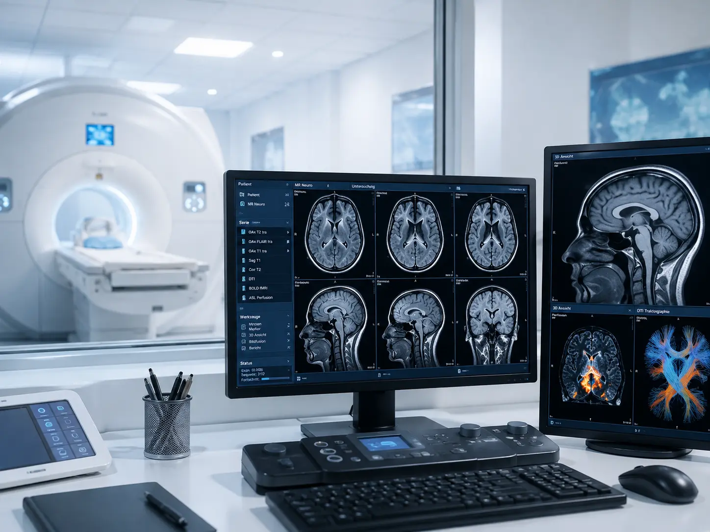
Diagnostic neuroradiology deals with the imaging examination of the central and peripheral nervous system — that is, the brain, spinal cord, and associated nerve structures. The primary methods used are magnetic resonance imaging (MRI) and computed tomography (CT), supplemented by specialized techniques such as vascular imaging (angiography) or functional imaging. The choice of method depends greatly on the urgency of the situation. In acute emergencies, such as suspected stroke, a computed tomography scan is often performed first. CT provides an initial, decisive diagnostic basis within minutes, allowing the treating physician to distinguish between a cerebral hemorrhage and a vascular occlusion. This distinction is critically important because it directly determines the treatment approach. While a vessel-opening therapy must not be carried out in the case of a hemorrhage, rapid reopening of the vessel can be life-saving in the case of a vascular occlusion.
Magnetic resonance imaging, in turn, is used when detailed visualization of nerve structures is required. It is particularly sensitive to early changes in the brain, such as small infarcts, inflammatory conditions like multiple sclerosis, or tumors. Spinal conditions — such as herniated discs or spinal cord damage — can also be precisely assessed with MRI. A key advantage is that MRI not only reveals structural changes but can also provide indications of inflammatory activity, circulatory disorders, or tissue damage.
In addition, specialized neuroradiological techniques can be used to visualize blood vessels — for example, in cases of suspected vascular stenosis, aneurysms, or vascular malformations. These examinations allow for a precise analysis of the condition of the vascular system in the brain and an assessment of the risks of stroke or bleeding.
Interventional Neuroradiology
Interventional neuroradiology extends diagnostic imaging to include therapeutic procedures performed minimally invasively via blood vessels. The goal is to treat diseases of the brain and its vessels directly, without the need for open surgery. These procedures are typically based on catheter technology: through an access point in the groin or arm, a thin catheter is introduced into the vascular system and advanced to the cerebral vessels under continuous X-ray guidance.
A particularly important area of application is the treatment of acute stroke. When a vascular occlusion is present, the blood clot can be mechanically removed through a procedure known as thrombectomy. A specialized instrument is guided through the catheter to the site of the occlusion in order to grasp and extract the clot. This treatment must be carried out as quickly as possible, since every minute without adequate blood flow can lead to irreversible brain damage. Interventional neuroradiology enables a targeted and often highly effective restoration of blood flow. Another key area of application is the treatment of aneurysms — pathological bulges in cerebral blood vessels that carry a high risk of dangerous bleeding. Fine platinum coils or special stents can be introduced into the aneurysm via the catheter to close it from the inside, significantly reducing the risk of rupture. Vascular malformations or certain tumor vessels can also be treated in this way. Throughout all of these procedures, continuous imaging provides precise control over the position of the instruments and the success of the treatment. The physician can determine immediately from the course of the procedure whether the vessel is patent again, whether an aneurysm has been safely occluded, or whether further measures are necessary. Interventional neuroradiology thus combines highly precise diagnostics with immediate therapy and is today a central component of modern stroke and vascular medicine.
All Experts in this Department
Show All
- Radiology and radiation therapy
Prof. Dr. Hendrik Wolff
Life Beam
- Radiology and radiation therapy
Prof. Dr. Marc Regier
Radiologie München im Alten Hof
- Radiology and radiation therapy
Prof. Dr. Christopher Herzog
Radiologie im Rotkreuzklinikum
- Radiology and radiation therapy