© PMC | AI
Nuclear Medicine
Nuclear medicine is an independent medical specialty closely related to radiology. While classical radiology primarily visualizes the structure of organs and tissues — such as their shape, size, and location — nuclear medicine focuses on function and metabolic processes within the body. It therefore answers not so much the question "What does this look like?" but rather "How does this organ or tissue work?" Nuclear medicine is used whenever functional information is essential, as it reveals how the body is actually functioning.
Scintigraphy & SPECT
Scintigraphy is a nuclear medicine procedure that depicts not the structure of organs, but their function and metabolic activity. To obtain this information, the patient is given a mildly radioactive substance — known as a tracer — by injection. The amount administered is so small that it does not interfere with bodily functions. The tracer is designed to accumulate selectively in certain organs or cell types, such as the thyroid, bones, or metabolically active tumor cells. As the tracer distributes throughout the body, it emits weak radiation that is picked up by a specialized camera. From these signals, the camera generates informative images showing how active specific tissues are.
Scintigraphy is primarily used when functional information is needed that cannot be adequately captured by conventional imaging techniques such as CT or MRI. A typical application is thyroid diagnostics, where it is used to assess whether individual areas are overactive or underactive. In cardiac diagnostics, it helps identify circulatory disorders of the heart muscle, and in the skeletal system, bone scintigraphy can provide evidence of inflammation, tumors, or metastases.
SPECT (Single Photon Emission Computed Tomography) is an advanced form of classical scintigraphy. It produces three-dimensional cross-sectional images, enabling the distribution of the radioactive substance to be localized with much greater spatial precision. This significantly improves diagnostic accuracy, particularly in the case of more complex findings. The results allow important conclusions to be drawn about the functional capacity of organs — for example, whether tissue is working normally, is overactive, or has been altered by disease. Scintigraphy therefore frequently provides crucial evidence of illness even before any structural changes become visible.
PET-CT / PET-MRI
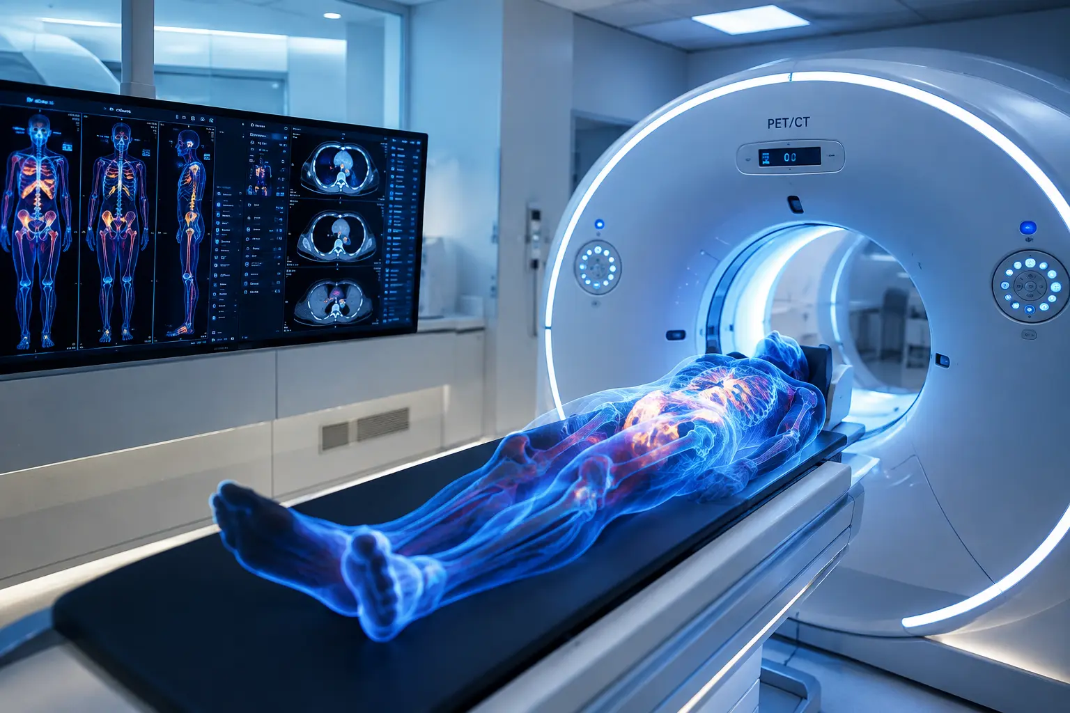
Positron Emission Tomography (PET) is a highly sensitive procedure for visualizing metabolic processes in the body. As with scintigraphy, a radioactive tracer is administered that accumulates preferentially in metabolically active cells. In oncology, a sugar analogue is frequently used, since tumor cells generally have an increased energy demand and therefore absorb more glucose than healthy tissue. PET makes this activity visible, revealing where in the body particularly active cellular processes are occurring.
PET is especially powerful when combined with computed tomography or magnetic resonance imaging, as this pairing brings together functional and anatomical imaging. While PET captures metabolic activity, CT or MRI provides precise spatial localization within the body. This makes it possible not only to detect that an abnormality is present, but also to determine exactly where it is located and what its structural characteristics are. The procedure is used particularly in cancer diagnostics, where it helps detect tumors, assess their extent, and distinguish between active tumor tissue and scar tissue. PET also plays an important role in treatment monitoring, as it can show at an early stage whether a therapy is working. In addition, PET is used in neurology — for example, in the investigation of dementia disorders or epileptic foci.
Radionuclide Therapy
Radionuclide therapy is a therapeutic procedure in nuclear medicine in which radioactive substances are used in a targeted way to treat disease. Unlike diagnostic applications, where only small amounts of radioactive material are used, therapeutically effective doses are administered here in order to selectively destroy pathologically altered tissue or to inhibit its function. A classic example is radioiodine therapy for thyroid disorders. Radioactive iodine is administered and accumulates specifically in the thyroid gland, owing to its natural capacity to store iodine. The radiation emitted acts locally, destroying overactive or diseased thyroid cells while largely sparing the surrounding tissue. Radionuclide therapy is also increasingly being used in oncology. Radioactive substances are coupled to molecules that bind specifically to tumor cells, delivering radiation directly to the site of disease while largely protecting healthy tissue.
The decision to proceed with radionuclide therapy is based on prior diagnostic examinations that confirm whether the tracer used accumulates sufficiently in the target tissue. During and after treatment, the response is monitored to assess how well the therapy is working and whether the target tissue has been successfully affected. These findings relate both to therapeutic success and to any potential side effects and the need for further treatment.
All Experts in this Department
Show All
- Radiology and radiation therapy
Prof. Dr. Hendrik Wolff
Life Beam
- Radiology and radiation therapy
Prof. Dr. Marc Regier
Radiologie München im Alten Hof
- Radiology and radiation therapy
Prof. Dr. Christopher Herzog
Radiologie im Rotkreuzklinikum
- Radiology and radiation therapy