© PMC | AI
Interventional Radiology
Vascular Interventions
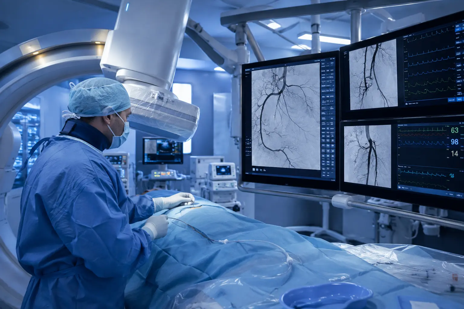
Vascular interventions are among the most important minimally invasive procedures in modern radiology. They are used to diagnose and simultaneously treat diseases of the arteries and veins. Many of these procedures are based on angiography — a specialized X-ray examination of the blood vessels in which contrast medium is injected through a thin catheter. The resulting images reveal whether a vessel is narrowed, blocked, or abnormally dilated. If a narrowing is identified — for example, in peripheral arterial occlusive disease (a chronic circulatory disorder of the extremities) or in narrowed coronary or renal arteries — treatment can often be carried out immediately within the same procedure.
Embolizations
Embolization is a non-surgical procedure in which blood vessels are deliberately blocked. The goal is to stop or significantly reduce blood flow in specific vessels. To accomplish this, special material is delivered through a catheter that is guided into the target vessel under fluoroscopic visualization, sealing it from the inside. Radiological imaging allows physicians to precisely identify the affected vessel and determine exactly how the embolization material needs to be positioned.
Tumor Interventions
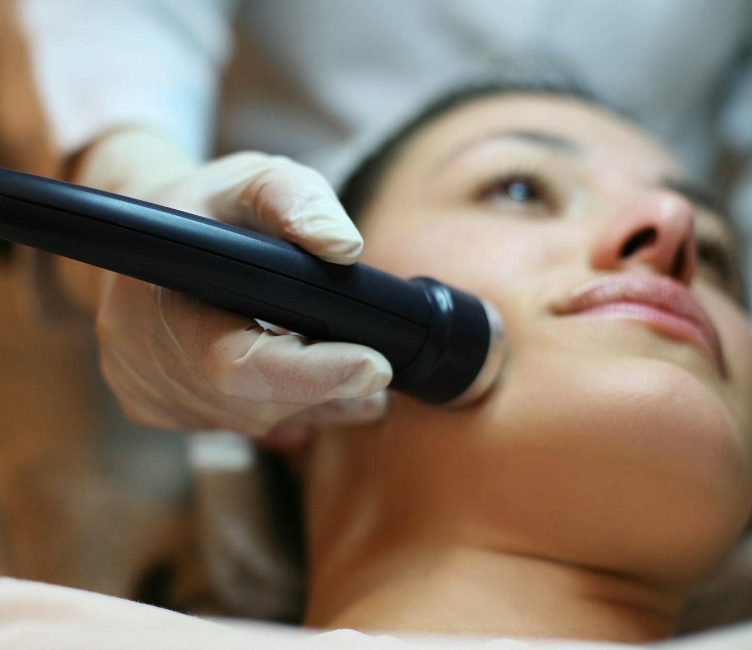
Tumor interventions are minimally invasive procedures for the local treatment of tumors, destroying the tumor directly within the body without the need for major surgery. One widely used approach is ablation. In this technique, a thin probe is inserted directly into the tumor under imaging guidance — most commonly CT, MRI, or ultrasound — and energy is then delivered through the probe to selectively destroy the tumor tissue. Depending on the technique used, this is achieved with either heat or cold. In radiofrequency ablation and microwave ablation, the tumor is destroyed through intense heating, whereas in cryoablation the tissue is cooled to a degree that causes the tumor cells to die. The choice of therapy ultimately depends on the size and location of the tumor. Imaging plays a critical role throughout this process: it not only shows the exact position of the tumor but also allows real-time monitoring during the procedure to confirm that the probe is correctly placed. After the procedure, imaging enables the physician to determine whether the tumor has been completely destroyed or whether further treatment is needed.
Image-Guided Biopsies & Drainages
Image-guided biopsies and drainages are important diagnostic and therapeutic procedures performed under ultrasound or computed tomography guidance. A biopsy is indicated when imaging has revealed an abnormality whose precise cause remains unclear — for example, a suspected tumor in the lung or liver. With accurate image guidance, the needle can be directed exactly to the suspicious site without injuring the surrounding tissue. The subsequent histological analysis of the extracted material provides essential information about whether the abnormality is benign or malignant and which course of treatment should be pursued. In the case of a drainage, imaging is used to monitor the correct removal of fluid collections from the body. Such collections occur, for example, in abscesses or develop in the context of inflammatory processes — arthritis being one example.
All Experts in this Department
Show All
- Radiology and radiation therapy
Prof. Dr. Hendrik Wolff
Life Beam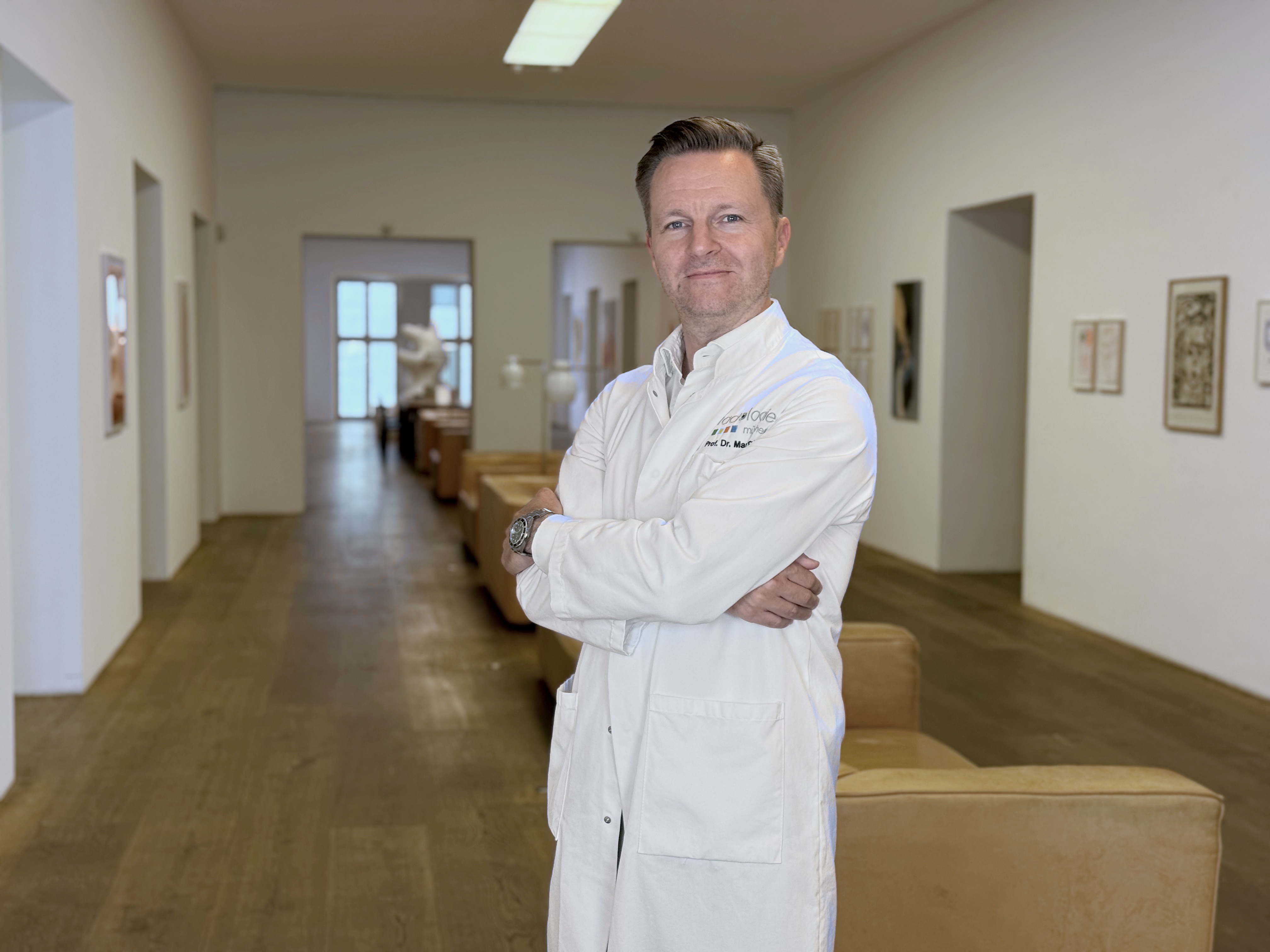
- Radiology and radiation therapy
Prof. Dr. Marc Regier
Radiologie München im Alten Hof
- Radiology and radiation therapy
Prof. Dr. Christopher Herzog
Radiologie im Rotkreuzklinikum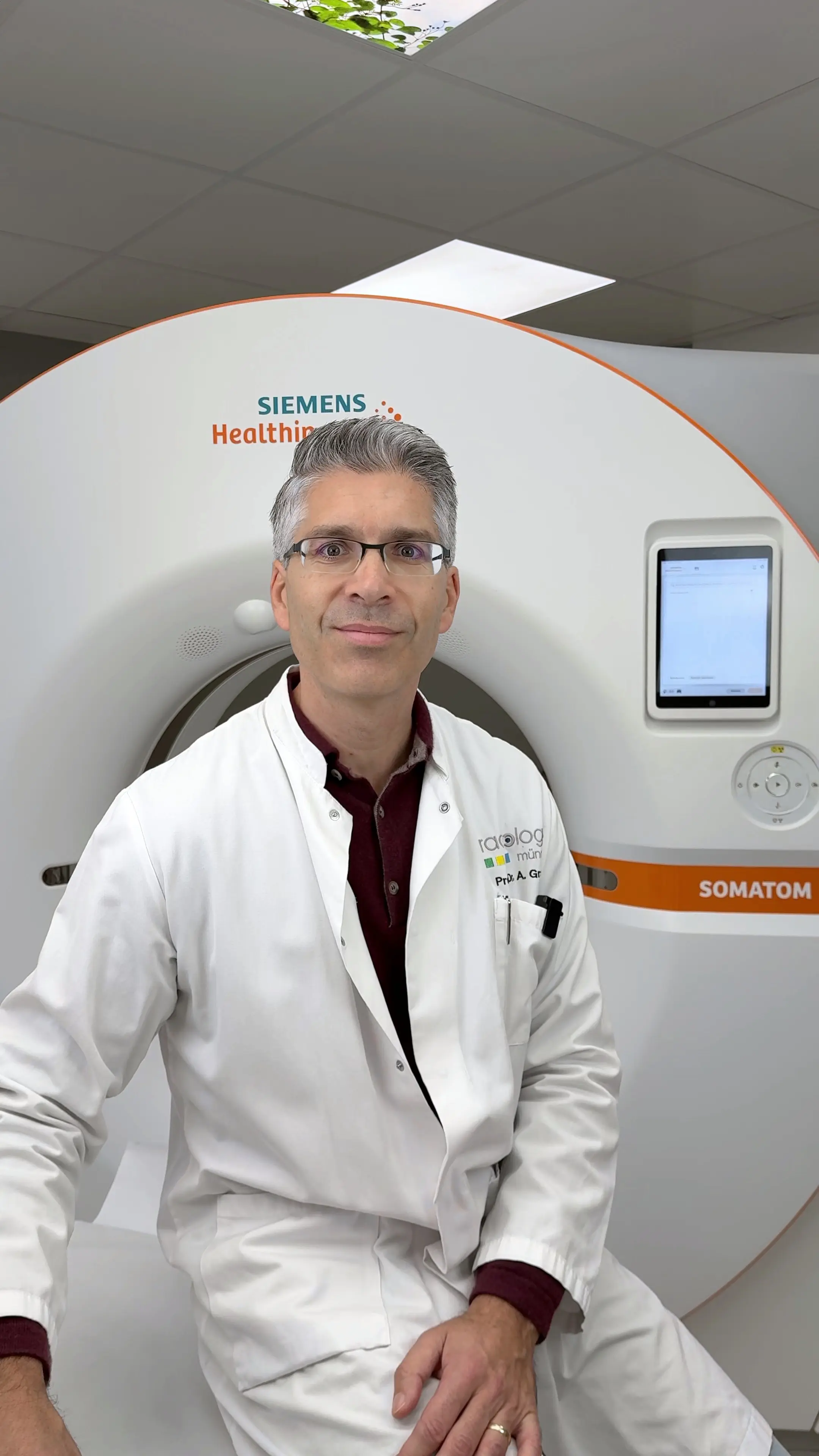
- Radiology and radiation therapy