.webp)
© PMC | AI
Magnetic Resonance Imaging (MRI)
MR Arthrography (Arthro-MRI)
MR arthrography, also known as arthro-MRI, is a specialized form of joint MRI in which a contrast agent is injected directly into the joint before imaging begins. The contrast agent distends the joint space and coats fine structures, allowing them to be visualized far more clearly than with conventional MRI.
The procedure is used primarily for complex diagnostic questions involving the shoulder, hip, wrist, or knee — for example, to evaluate cartilage, the joint lip (labrum), or fine ligament structures. Injuries that are difficult to detect on standard MRI, such as small labral tears or superficial cartilage damage, can be depicted with greater precision using MR arthrography. This makes it a particularly valuable basis for clinical decision-making, especially in sports medicine and ahead of planned joint-preserving procedures.
Whole-Body MRI
Whole-body MRI allows the entire body to be examined in a single session, without the use of ionizing radiation. The head, neck, chest, abdomen, pelvis, and extremities are imaged in successive sections and then combined into one comprehensive picture. In this way, many organs, the nervous system, blood vessels, the skeleton, and soft tissues can all be assessed in a single examination.
Whole-body MRI is used, among other things, in oncological diagnostics — for example, to search for metastases or to monitor the course of certain cancers — as well as in inflammatory and rheumatic diseases that can affect multiple regions of the body. The procedure is also growing in importance as a preventive tool, since it can detect structural abnormalities across different body regions without exposing the patient to radiation. Its diagnostic value depends heavily on careful selection of the clinical question and experienced interpretation of the findings.
Experts for this Treatment Method
All Experts in this Department
Show All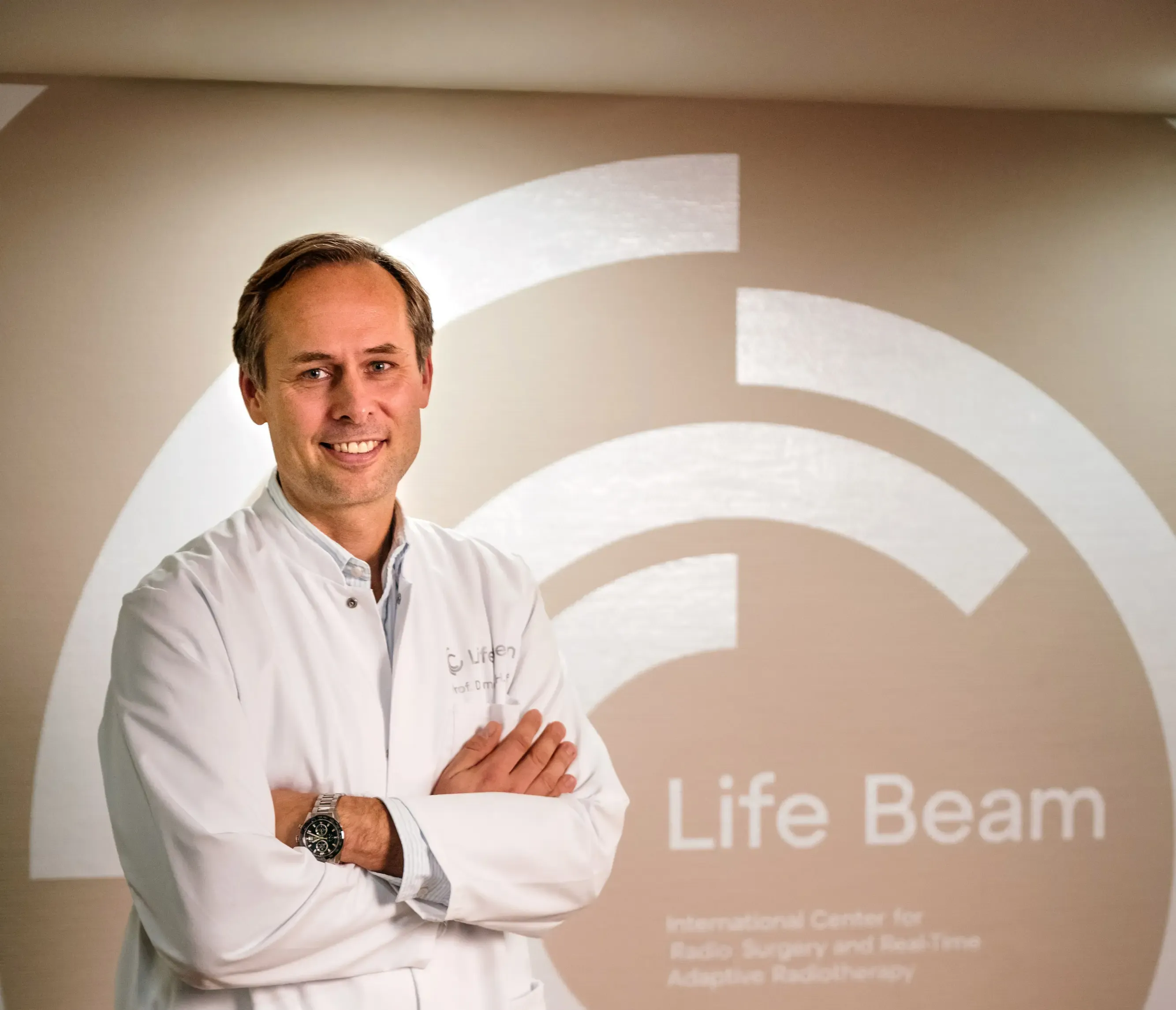
- Radiology and radiation therapy
Prof. Dr. Hendrik Wolff
Life Beam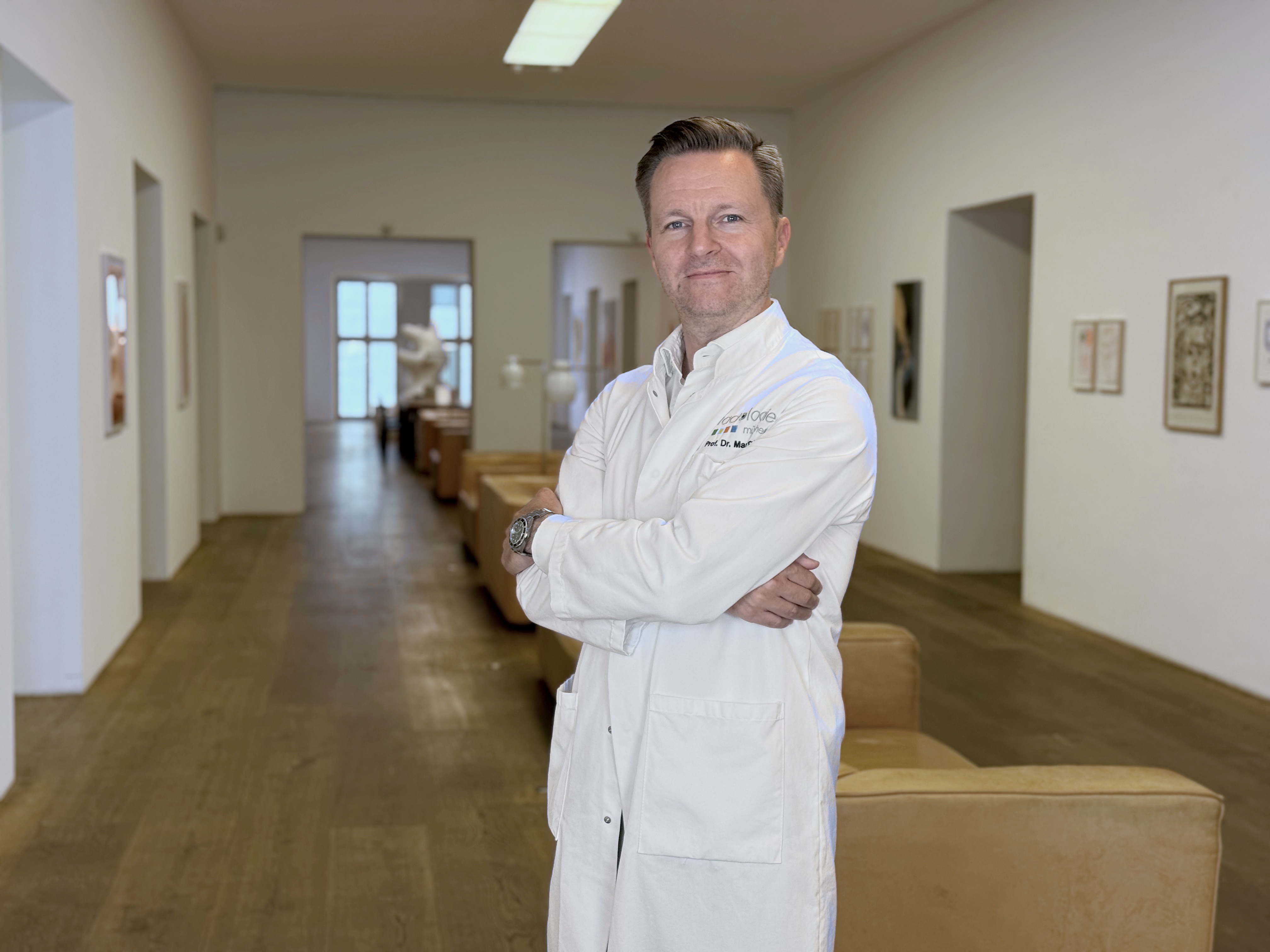
- Radiology and radiation therapy
Prof. Dr. Marc Regier
Radiologie München im Alten Hof
- Radiology and radiation therapy
Prof. Dr. Christopher Herzog
Radiologie im Rotkreuzklinikum
- Radiology and radiation therapy