© PMC | AI
Cardiac Imaging
Cardiac CT (Cardio-CT)
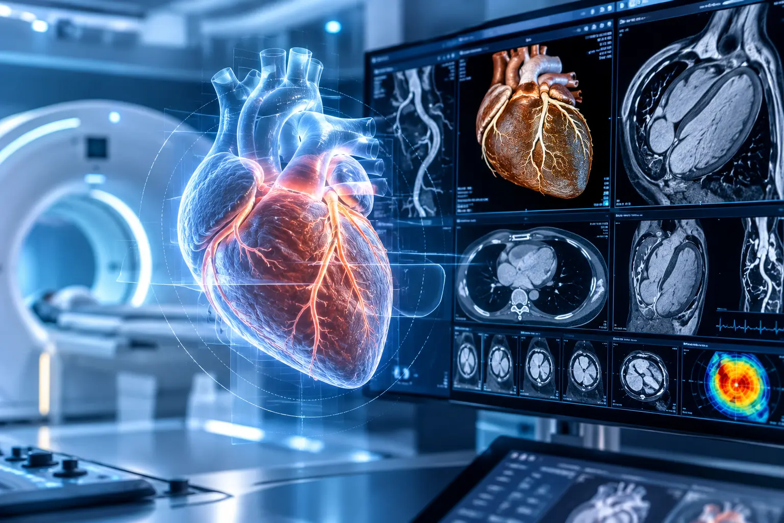
Cardiac CT, also known as cardio-CT, is a state-of-the-art procedure for examining the coronary arteries and is used primarily for the early detection of coronary artery disease. Unlike conventional methods such as stress ECG or scintigraphy — which only reveal circulatory problems once cardiac function is already significantly impaired — computed tomography can detect abnormal deposits and narrowings in the coronary arteries at an early stage, often long before any symptoms develop.
Cardiac CT is indicated, for example, in cases of unexplained chest pain or shortness of breath, as well as in individuals at increased risk of coronary artery disease, such as those with diabetes, high blood pressure, or a family history of the condition. Beyond the coronary arteries, the procedure can also assess the heart muscle, heart chambers, and heart valves, enabling a wide range of cardiac conditions — including vascular narrowings, valve diseases, and congenital heart defects — to be diagnosed with minimal invasiveness. The examination is performed with contrast medium and is rendered nearly artifact-free through synchronization with the heartbeat, a process known as ECG triggering. Compared to invasive cardiac catheterization, cardiac CT is considerably gentler for most patients: no anesthesia is required, the radiation dose can be kept low, and there is no risk of vascular injury.
Cardiac MRI (Cardiac MRI)
Magnetic resonance imaging of the heart complements cardiac imaging by providing information that goes beyond the visualization of blood vessels alone. It uses no ionizing radiation and delivers detailed data on heart muscle function, pumping capacity, blood flow, and any scarring or inflammatory changes in the tissue. This makes cardiac MRI particularly valuable for determining the cause of heart failure, assessing the extent of a previous heart attack, or identifying inflammation of the heart muscle.
While cardiac CT focuses primarily on imaging the coronary arteries, cardiac MRI addresses how well the heart muscle itself is functioning and whether — and to what degree — tissue has been damaged. The findings can guide critical treatment decisions, such as whether medication, an interventional procedure, or surgery is the most appropriate course of action. Both methods complement each other and are used individually or in combination depending on the clinical question at hand.
Experts for this Treatment Method
All Experts in this Department
Show All
- Radiology and radiation therapy
Prof. Dr. Hendrik Wolff
Life Beam
- Radiology and radiation therapy
Prof. Dr. Marc Regier
Radiologie München im Alten Hof
- Radiology and radiation therapy
Prof. Dr. Christopher Herzog
Radiologie im Rotkreuzklinikum
- Radiology and radiation therapy