© Maks Gelatin
October 15, 2026
Christine Bürg & Marianne Waldenfels
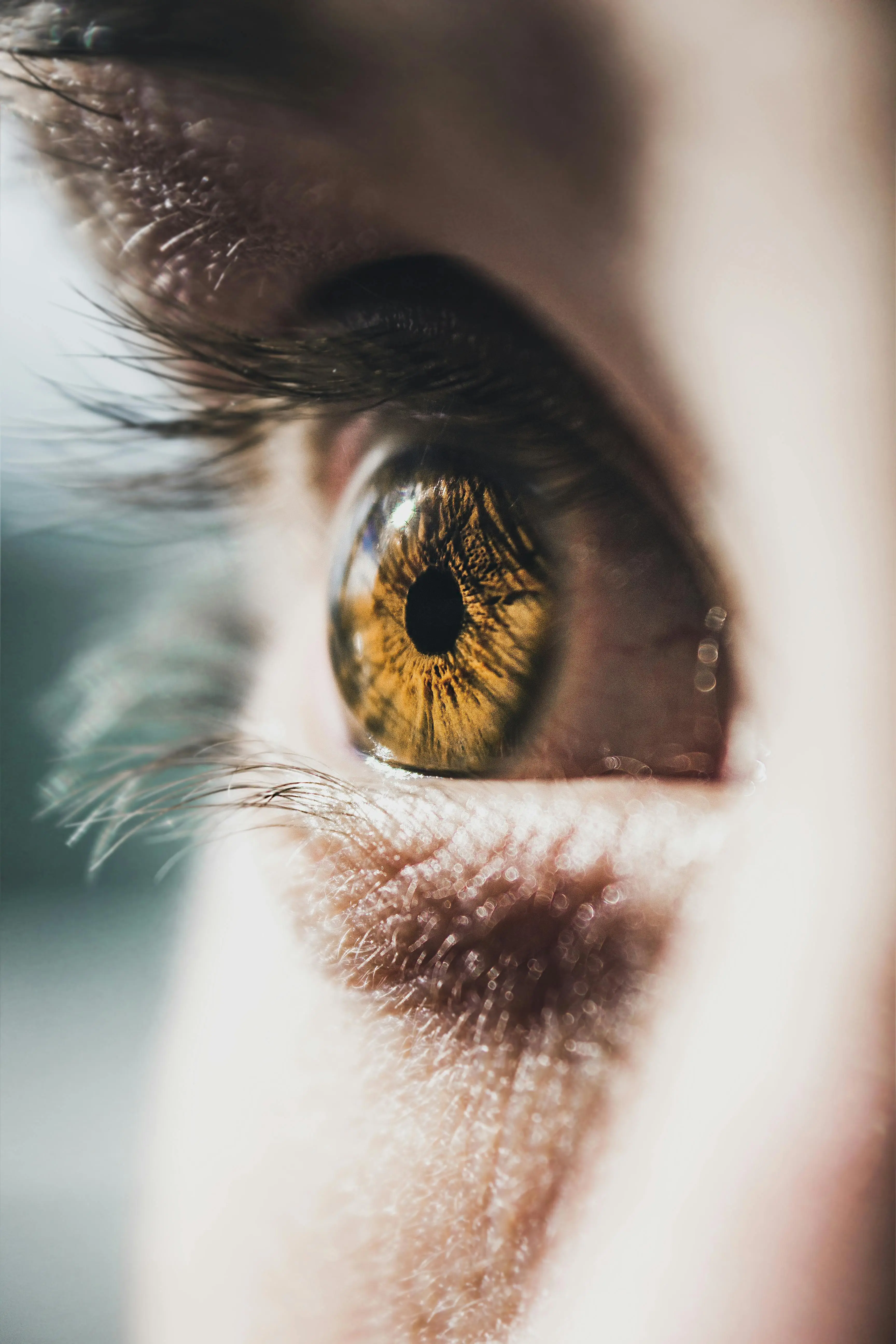
Almost everyone has seen them, and most people ignore them: tiny specks and strands drifting across their vision. Usually, they’re harmless. But sometimes they signal an emergency where quick action can save your sight. Ophthalmologist Dr. Mirka Höltzermann explains which warning signs to watch for.

An interview with
Dr. Mirka R. Höltzermann
While reading, gazing at the sky, or looking at a screen, they suddenly appear: tiny shadows that follow every eye movement and vanish the moment you try to focus on them. Specialists call them mouches volantes — French for "flying gnats" — or, in English, floaters. They are usually caused by a natural change in the vitreous body. However, if the shadows appear for the first time or are accompanied by flashes of light, they may indicate a retinal tear or the onset of a retinal detachment.
The problem: many people wait too long. By the time they finally see a doctor, the retina has often already detached. In an interview, Dr. Mirka Höltzermann, a specialist in ophthalmology, explains how to tell harmless vitreous opacities apart from genuine warning signs, who is at increased risk for retinal problems, and what laser treatment and surgery can do about bothersome floaters.
Dots, threads, and streaks drifting through your field of vision: what exactly are mouches volantes?
The eye is filled with a gel-like substance called the vitreous body, which consists of water and collagen fibers. As we age, the vitreous shrinks and the collagen fibers clump together. This then becomes visible as threads or flies — mainly when looking at white walls or a bright screen.
Why do mouches volantes become more common with age?
As mentioned, the older we get, the more the vitreous shrinks — and its water content decreases. This leads to increasing opacities. These flecks or flies can be the first sign of a posterior vitreous detachment, in which the vitreous separates from the retina. This process can cause damage such as holes or tears in the retina, which may lead to a retinal detachment — a true ophthalmic emergency.
A new Dutch study examined which symptoms indicate a retinal tear or retinal detachment. What does this mean for people who suddenly notice new spots in their visual field?
When new flies or flecks appear, it is advisable to have a retinal check with an ophthalmologist promptly. Even without additional warning signs, the development of a retinal tear or retinal detachment is not unlikely.

For which symptoms should you not wait for a routine appointment?
Flashes of light, a large number of black dots/a shower of soot, and a curtain-like shadow in your vision are alarm signals that something is wrong with the retina. In these cases, an emergency visit to an ophthalmologist is indicated — on Sundays or public holidays, this means going to the ophthalmological emergency service.
Who is particularly at risk: do nearsightedness, previous eye surgery, or a family history play a role?
Yes, there are patients with a higher risk of vitreous detachment and retinal detachment. These include people who are nearsighted. In these individuals, the vitreous tends to shrink earlier and the retina is usually thinner, meaning that when the vitreous detaches it can cause retinal holes or tears and a retinal detachment. Patients who have undergone eye surgery, patients with a positive family history, and patients who have experienced trauma to the eye — for example, from a tennis ball or a champagne cork — are also at higher risk.
Is there anything you can do yourself to prevent retinal detachment?
Absolutely. I discuss the increased risk with nearsighted patients and recommend regular retinal check-ups, as well as after surgery on the eye — even at a young age following refractive procedures. I also think it is important to exercise a certain level of caution during sports, when opening corked bottles, and when doing DIY work.
If the first symptoms of a vitreous detachment or retinal defect appear, it makes sense not to wait. Unfortunately, many patients still come to the practice too late, and the retina has already detached.
What exactly do you examine when someone comes to you with spots that have appeared suddenly in their visual field?
In general, I always examine the entire eye — as well as the fellow eye — using a slit lamp, which is a specialized microscope. This is followed by funduscopy, meaning an examination of the fundus of the eye using a lens with the pupil dilated by drops. This allows the entire retina, including the peripheral areas, to be visualized.
A wide-field photograph of the retina and a Spectralis OCT (optical coherence tomography) of the central retina are also very helpful. The vitreous and retina are examined for signs of detachment. In the vitreous, these may include flecks or "flies" or what is known as a Martegiani ring; in the retina, they may include bleeding, holes, or even a detachment of the retina.
Laser treatments are offered for bothersome floaters, but there are also reports of risks. How do you assess these procedures, and what alternatives are available?
Laser vitreolysis for vitreous floaters can be a worthwhile option. What matters is the nature of the floater — whether it is clearly defined or more of a streaky change across the various levels of the vitreous — and the degree of distress it causes the patient. If they have found the floaters very limiting and disruptive for more than six months, then laser treatment may be appropriate.
In phakic patients — that is, patients who have not yet had cataract surgery — the laser can damage the lens to the point where it becomes cloudy. In addition, multiple laser sessions are usually required. I always conduct a risk-benefit analysis with my patients.
If the distress is extreme and the floaters are very dense and central and have persisted over a long period of time, a vitrectomy — that is, the surgical removal of the vitreous — can be discussed after the most careful risk-benefit analysis. There are also dietary supplements containing micronutrients that are intended to prevent further clumping of the collagen fibers and restore the balance within the vitreous.

Almost everyone has seen them, and most people ignore them: tiny specks and strands drifting across their vision. Usually, they’re harmless. But sometimes they signal an emergency where quick action can save your sight. Ophthalmologist Dr. Mirka Höltzermann explains which warning signs to watch for.
Christine Bürg & Marianne Waldenfels

An interview with
Dr. Mirka R. Höltzermann

Can fasting relieve hot flashes and other menopausal symptoms? Dr. Verena Buchinger-Kähler shares remarkable cases from her clinical practice and explains how women respond to prolonged fasting – and what science has revealed so far.
Christine Bürg & Marianne Waldenfels
An interview with
Dr. med. Verena Buchinger

"Feel good, live better" – in this episode, Dr. Alexandra Schoeneich sheds light on the wide-ranging changes that menopause brings to the body
Christine Bürg

An interview with
Dr. med. Alexandra Schoeneich

Stopping antidepressants can trigger symptoms that resemble a return of depression. Psychiatrist Prof. Dr. Andreas Menke explains how to tell the difference, why the final milligrams can be particularly difficult, and which two mistakes patients should avoid.
Christine Bürg & Marianne Waldenfels

An interview with
Prof. Dr. med. Andreas Menke