Other Treatment Methods
.webp)
Laser eye surgery (Refractive surgery)
Refractive surgery unites various treatment methods. They all share the common goal of eliminating the need for patients to use visual aids like glasses or contact lenses. Refractive errors are not to be understood as a disease. They are rather the result of an individual deviation in the design of the optical system "eye". This results in refractive errors. To correct them, the eye surgeon takes advantage of the fact that the refraction of light at the cornea and lens is very strong at 45 and 22 diopters, and even the smallest changes in these refractive powers can have a significant effect on the entire optical system.
Before laser eye surgery
Treatments within the framework of refractive surgery are not considered absolutely necessary operations on a healthy eye from the perspective of the ophthalmologist. A comprehensive diagnosis of the entire eye is therefore an important basis for preparation and consultation. Based on the findings obtained, the doctor and patient together select a suitable method to correct the refractive error. Nowadays, there are two different treatment concepts available for this purpose.
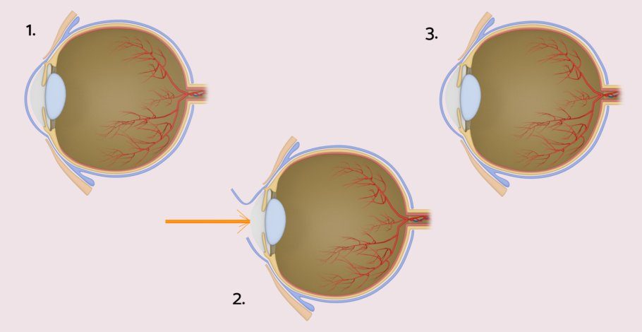
Laser treatment of the cornea
The probably best-known method for correcting a refractive error is laser treatment of the callus. Here, the eye surgeon modulates the curvature, and thus the refractive power of the cornea, by removing corneal tissue and thereby correcting the existing refractive error (myopia or farsightedness and/or astigmatism). Alternatively, they can implant a special lens in addition to the human lens to correct the visual impairment.
Eye laser: The LASIK procedure
In the meantime, numerous acronyms exist for the different laser treatments. However, they all basically work on the same principle: The slight change in the curvature of the strongest refractive medium, the human cornea, effectively changes the entire optical system. The most commonly performed LASIK technique worldwide consists of two steps. First, the ophthalmic surgeon prepares a superficial, thin (approximately 120 μm) corneal flap, which has a diameter of about 9 mm. For this, they use a microkeratome, an extremely small cutting tool for eye laser treatments, or the femtosecond laser. Subsequently, they treat the exposed deeper corneal tissue with an excimer laser, based on the data from preliminary examinations. During this process, they vaporize tissue and thus change the curvature of the cornea. Then they reposition the previously prepared corneal flap onto the wound surface. The body's own tissue ensures rapid wound healing with low pain and quick restoration of vision.
Risks of eye laser surgery
Both concepts carry the risk of wound surface infection (laser treatment) or intraocular infection (lens implantation). In rare cases, a minor visual error may still persist after treatment, which the eye specialist can usually correct in a second treatment step.
After the eye laser surgery
In the first few days after laser treatment, the patient may experience mild pain and discomfort in the form of foreign body sensation, light sensitivity, and still blurry vision. The same applies to the implantation of an intraocular lens. A significant difference between the two procedures is that laser treatment is performed on both eyes on the same day, whereas lens implantation takes place on different days. There should be about a week of recovery time in between. Patients can quickly resume their daily activities after both procedures. Ball sports, swimming, etc., should be avoided for up to two months after the surgery.
Other Specializations
Experts for this Treatment Method
- Modern Ophthalmology
Dr. med. Ilya Kotomin
Smile Eyes Leipzig
- Modern Ophthalmology
Priv.-Doz. Dr. med. Daniel Pilger
Smile Eyes Berlin
- Modern Ophthalmology
Raphael Neuhann (FEBO)
Opthalmologikum Dr. Neuhann / Augentagesklinik am Marienplatz
- Modern Ophthalmology
Dr. med. Tabitha Neuhann
Opthalmologikum Dr. Neuhann / Augentagesklinik am Marienplatz
- Modern Ophthalmology
Prof. Dr. med. Tanja M. Radsilber
Augenzentrum Prof. Dr. med. Holzer & Prof. Dr. med. Rabsilber
- Modern Ophthalmology
Dr. med. Karsten Klabe
Breyer, Kaymak & Klabe AugenchirurgieAll Experts in this Department
Show All
- Modern Ophthalmology
Dr. Christine Heun
IRISIOvital Praxis für Augengesundheit
- Modern Ophthalmology
Dr. Mirka R. Höltzermann
Augenpraxis Dr. Höltzermann, Dr. von Schnakenburg, Augenpraxis Dres. Höltzermann & von Schnakenburg- Modern Ophthalmology
Dr. med. Ilya Kotomin
Smile Eyes Leipzig- Modern Ophthalmology
Priv.-Doz. Dr. med. Daniel Pilger
Smile Eyes Berlin- Modern Ophthalmology
Raphael Neuhann (FEBO)
Opthalmologikum Dr. Neuhann / Augentagesklinik am Marienplatz- Modern Ophthalmology