Other Treatment Methods
- 4D static analysis and dynamic pedography
- Acupuncture
- Arthrosis therapy
- Artificial elbow joint
- Artificial hip joint
- Artificial knee joint
- Artificial shoulder joint
- Bioregenerative Orthopedics
- Endoprosthetics
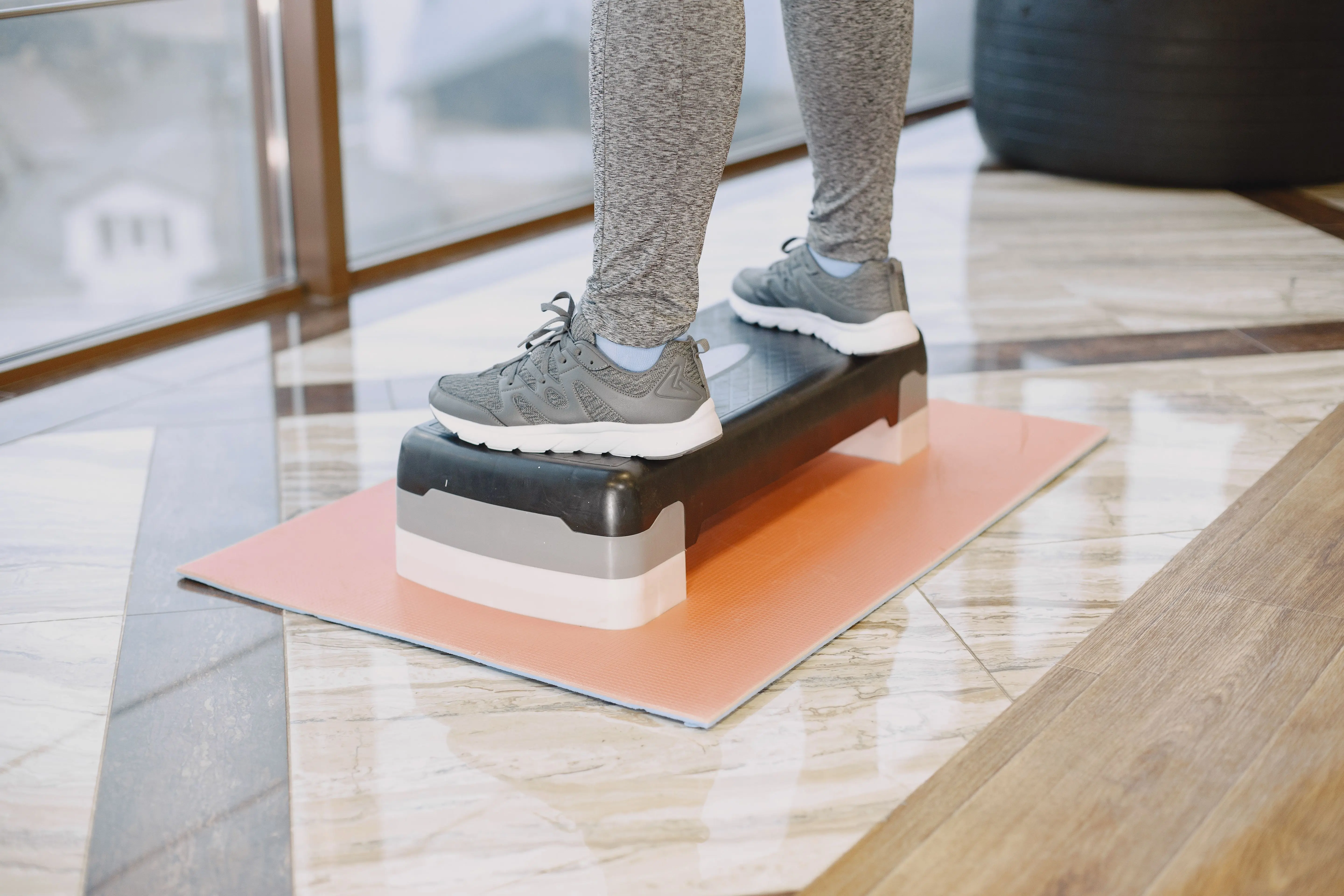
- Medical training therapy
- Open MRI and MRI-guided injection therapy
- Orthopedic pain therapy
- Osteopathy
- Shock wave therapy
- Sports orthopedics / sports medical treatment

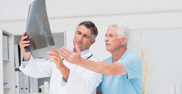
Artificial shoulder joint
The shoulder joint occupies a special position in the human body: it allows the greatest range of motion of all joints. To achieve this, a spherical joint head pairs at the upper end of the upper arm with a comparatively shallow socket on the shoulder blade. To ensure both mobility and stability, and to securely center the head of the upper arm in the joint socket, the joint has various mechanisms:
– the surrounding muscle cuff,
– a cartilaginous labrum, and
– an (atmospheric) negative pressure in the joint
A shoulder joint only functions perfectly in the long term if all components are in good working condition. Injuries such as a bone fracture or a dislocation of the shoulder joint can lead to long-term consequential damages and thus make an artificial joint necessary. But the modern, sedentary lifestyle also has consequences for the shoulder's function. Frequent sitting leads to a poor posture of the upper body and thus changes the tension conditions of the shoulder girdle muscles. The rotator cuff is often also defective as a sign of pre-arthrosis. In Germany, the prevalence of this in men beyond the age of 70 is around 30 percent. With a weakened rotator cuff, the head of the upper arm tends to rise. This paves the way for later shoulder joint arthrosis (omarthrosis). Depending on the severity, the arthrosis leads to severe pain and massive restrictions in movement that impair everyday life.
Infections or tumors rarely make shoulder joint replacement necessary. Tumors Only in the past decade has shoulder endoprosthetics become routine – later than hip- and knee joints. Meanwhile, specialists in Germany implant around 60,000 shoulder endoprostheses annually, with a rising trend. As with the replacement of other major joints, it is assumed that an artificial shoulder joint lasts about 15 years.
Models of prostheses for the shoulder
In cases of partial or complete replacement of the shoulder joint, various systems are possible. Both partial replacement (hemi-prosthesis, only the joint surface of the humeral head) and total endoprosthesis (TEP) with head and socket have their proven areas of application. The choice of prosthesis depends not only on the joint damage but also on the condition of the surrounding tissue (rotator cuff), the patient's age, and the quality of the bone.
Total hip arthroplasty (THA)
During shoulder THA, orthopedists use anatomical systems for one. Both the head and socket are replaced when both joint surfaces are severely affected. The musculature, i.e., the rotator cuff, must be intact for this prosthesis. Because long prosthesis shafts complicate the later replacement of a prosthesis, the trend is towards using shorter shafts.
Numerically dominated by older patients — and this patient age group is the largest — reverse systems, a peculiarity of shoulder arthroplasty, dominate. Here, the head and socket are "switched." The head is fixed to the scapula and the socket to the humerus. This type of shoulder prosthesis is oriented towards the damage pattern and not the original anatomy of the shoulder. The results of these surgeries are good, even if the arthritis is advanced or the surrounding muscle cuff of the shoulder joint is no longer intact.
Additionally, orthopedists use special fracture prostheses, for example, when it has not been possible to reconstruct the shoulder joint from fragments in the case of a complicated humerus fracture.
Partial prosthesis
During the implantation of a hemi or partial prosthesis, significant parts of the joint are preserved: The socket remains intact, and the joint-surrounding muscles are left undamaged. This allows for early rehabilitation in which everyday movements can be practiced again. Meanwhile, there are so-called cup prostheses that do completely without a shaft and only replace the joint surface. The cup prosthesis is screwed into the head of the humerus. In addition to solely replacing the humeral joint surface, it can also be combined with a prosthesis of the shoulder socket.
Cemented or uncemented
Prosthetics are implanted cemented, uncemented (“press-fit”, ingrowing), and/or screwed. The choice of fixation depends, among other things, on the quality of the bones.
Whenever possible, the surgeon aims for cement-free fixation of the prosthesis. The advantage is that no foreign material is required apart from the prosthesis. The surfaces of cement-free prostheses are slightly roughened, encouraging the bone to grow around them. The connection between bone and prosthesis is more natural than when using cement. In the reverse prosthesis, the new joint head is always implanted without cement (screwed). Even small prostheses like the cup prosthesis do not require cement.
For the procedure of cement-free fixation, the quality of the bone must be good. If bone density and strength are reduced due to osteoporosis or necrosis, the surgeon will cement the prosthesis. A special two-component acrylic resin is used as bone cement.
Operative approach
For most shoulder prosthesis implantations, an incision about ten centimeters long is made in the anterior area of the shoulder, often between the deltoid muscle (outer) and the large pectoral muscle (inner). Less commonly, the surgeon uses lateral approaches, in which the large shoulder or deltoid muscle is opened (“Delta-Split”).
Minimally invasive procedure
In many areas of surgery, minimally invasive procedures established with “keyhole surgery”. In endoprosthetics one of the stated goals is to traumatize the tissue as little as possible. Also, from a cosmetic point of view, minimally invasive approaches to the shoulder joint are not insignificant. However, when inserting prostheses, the surgeon is limited when using minimally invasive approaches: he must be able to fully view and, if necessary, completely remove the joint parts. Exclusively minimally invasive techniques are therefore reserved for partial joint replacement at the shoulder. Nevertheless, the improved surgical technique has led to smaller access routes. As a result, the surgeon spares the muscles, tendons, cartilage surfaces and bones of the shoulder and proceeds as little traumatizing as possible. However, certain steps are sometimes unavoidable: For larger prostheses (hemiarthroplasty, TEP, inverse prosthesis), the rotator cuff must be partially severed and then sutured again.
The procedure
The procedure usually takes place under general anesthesia. Spinal anesthesia, where the patient remains awake, is possible upon request or with certain accompanying illnesses. First, the patient is placed in a semi-seated position (“beach chair position”). The skin is disinfected and the incision is made depending on the prosthesis used. To keep an eye on the joint, the surgeon must cut through muscles. He acts gently and protects the surrounding tissue. He then prepares the bone joint for prosthesis implantation. During the implantation itself, it is advantageous for the tissue that most shoulder prostheses are modular. This means that the access for the individual components can be kept as small as possible. In addition, only individual parts of the prosthesis can be replaced. After insertion, the prosthesis is checked for a firm, correct fit and the shoulder joint for mobility while still in the operating room. In 80 to 90 percent of cases, the shoulder prosthesis results in pain-free function and good mobility sufficient for everyday life.
Navigation systems in shoulder endoprosthetics
Crucial for a long-term satisfactory result in the implantation of a shoulder prosthesis is the correct fit. The shoulder joint, with its large range of motion, presents particular challenges for the surgeon. The perfect position of the prosthesis parts is therefore individually adjusted for each patient. As shoulder endoprosthetics have evolved, implantation techniques have become increasingly refined, thereby avoiding earlier causes of loosening. Modern three-dimensional navigation systems can make it easier for the surgeon to find their way around the surgical area on the shoulder and ensure that the prostheses fit more accurately.
Risks of shoulder endoprosthetics
The renowned journal Lancet referred to joint replacement as the 'operation of the 21st century' in 2007. Today, prostheses usually last around 15 to 20 years, often longer. These numbers are steadily climbing: Between 1993 and 2003, the percentage of hip replacement prostheses that were functioning well after 10 years increased from 92% to 95%. Besides prosthetic longevity, the knowledge of potential complications is crucial for the long-term evaluation of an artificial joint's success. The most serious risks of joint replacement surgery include infections (early, late; superficial, deep), formation of blood clots in a leg vein (thrombosis, possibly embolism), and so-called aseptic prosthetic loosening (without infection).
Crucial for prognosis in individual cases is the earliest possible detection of a complication and its immediate, appropriate treatment. Infections require treatment with possibly specifically selected antibiotics. In cases of deep infections, partial or complete removal of the prosthesis may be necessary (see below). Thromboses are detected by ultrasound examinations of the blood vessels and treated with medications that reduce the blood's clotting ability. In Germany, approximately 24,000 artificial hip and about 16,000 artificial knee joints are replaced annually. In about half of the cases, an infection is the underlying cause; furthermore, aseptic loosening (loosening without infection) and other mechanical causes are responsible for the prosthesis replacement. The DGOOC (German Society for Orthopaedics and Orthopaedic Surgery) has certified surgical centers (www.endocert.de) that ensure experienced surgeons oversee all procedures, all steps are documented, and the staff is regularly trained. The case numbers of these centers are publicly accessible.
For a better result of shoulder prosthesis
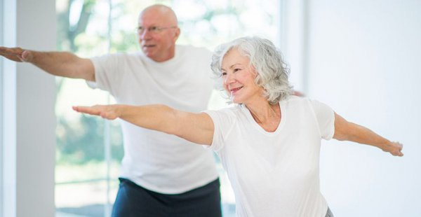
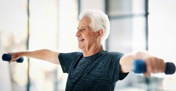
After undergoing an artificial joint replacement, pain-free movement should ideally be possible without crutches within six to eight weeks, at the latest after three months. Well-dosed sports activities are usually not a problem (hiking, walking, golf, cycling, swimming, strength training, etc.). However, artificial joints are not sports joints and should be treated with care and prudence. The better this is achieved, the higher the chances that an artificial joint will last for many years, ideally a lifetime. Modern high-tech implants and new surgical techniques help in this regard. Therefore, the patient's behavior and lifestyle are also very crucial for the treatment success. Weight control, strength building, coordination training (gait) as well as the type and extent of daily use are factors solely in the hands of the patient.
Prehabilitation
Preparation for a joint replacement surgery is not solely in the hands of the attending doctors. The patient can start preparations even before the procedure to facilitate the surgeon’s work and achieve a better outcome. Weight loss, muscle strengthening, gait training, and optimizing cardiovascular and metabolic values in consultation with the general practitioner ensure faster surgeries, shorter anesthesia time, fewer complications, a shorter and more successful follow-up treatment, and a faster recovery. General practitioner ensure faster surgeries, shorter anesthesia time, fewer complications, a shorter and more successful follow-up treatment, and a faster recovery.
Rehabilitation
A several-week intensive rehabilitation begins immediately following the hospital stay (usually one week after the surgery). It aims to ensure a continuous healing process and a goal-oriented development of the patient’s motor, cognitive, and psychological abilities. Pain is treated, wound healing is monitored, muscles are stabilized, coordination is trained, and self-confidence is rebuilt. Health status, home care, and potential complications determine whether a patient undergoes rehabilitation in an inpatient form (stay in a rehabilitation clinic with full care and therapeutic applications) or an outpatient form (living at home, treatments in one or more therapeutic practices). The hospital’s social service takes over the application for rehabilitation in coordination with surgeon and patient, and the health insurance covers the costs.
After shoulder prosthesis surgery
The surgery is followed by a relatively long rehabilitation phase. Patients should expect around six weeks of significant immobilization (wearing a shoulder brace) and up to six months before they can use their shoulder as usual. The reasons for the duration are the large range of motion to be achieved and the time-consuming development of the shoulder muscles, which are crucial for joint guidance. Patients usually wear a shoulder brace for four to six weeks, in which the affected arm is fixed in the so-called neutral position - upper arm vertical, slightly abducted, forearm horizontal.
Passive physiotherapy and mobilization of the shoulder joint performed by a specialist starts the day after the operation. This early promotion supports muscle development, coordination, and mobility in the shoulder joint. To ensure that the stitches in the skin, tendons, and muscles heal securely, active movements are prohibited in the first six weeks post-operation, and even passive movement range is limited.
Following the hospital stay, a three-to-four-week follow-up treatment takes place. Rehabilitation can be inpatient or outpatient. In outpatient rehabilitation, the patient’s social environment must provide support. For example, a shoulder patient cannot dress or undress, wash, or drive alone while wearing a brace. Most everyday movements like brushing teeth and dressing are possible again about two months post-operation. Continuing physiotherapy and independently performing the learned muscle strengthening and movement exercises at home is essential even after the follow-up treatment. Particularly building a functioning shoulder muscle structure that secures the prosthesis position takes many weeks, especially if the patient has suffered from pain for a long time. The patient’s initiative is required throughout the process, but must be appropriately regulated by the doctor and therapist. Many fitness studios now offer suitable training devices that can and should be used after medical supervision has ended. Competent guidance from the staff is essential.
Experts for this Treatment Method

- Innovative Orthopedics & Sports Orthopedics
Dr. med. Martin Volz & Kollegen
Sportklinik Ravensburg
- Innovative Orthopedics & Sports Orthopedics
Prof. Dr. Hans-Georg Simank
Zentrum für Orthopädie und Neurochirurgie
- Innovative Orthopedics & Sports Orthopedics
Prof. Dr. med. Sebastian Siebenlist
Sektion Sportorthopädie TU München, Klinikum rechts der Isar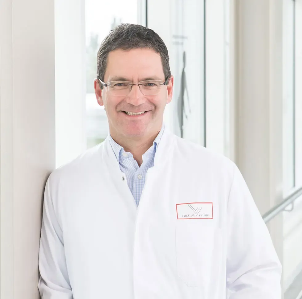
- Innovative Orthopedics & Sports Orthopedics
PD Dr. Wolfgang Pötzl
Vulpius Klinik Bad Rappenau
- Innovative Orthopedics & Sports Orthopedics
Prof. Dr. med. Christoph Lohmann
Orthopädische Universitätsklinik MagdeburgAll Experts in this Department
Show All- Innovative Orthopedics & Sports Orthopedics
Niklas Haberstroh
MW - Praxis für Orthopädie & Sportmedizin
- Innovative Orthopedics & Sports Orthopedics
PD Dr. med. Daniel P. Berthold
OrthoCenter München
- Innovative Orthopedics & Sports Orthopedics
Dr. med. Florian Pfalzer
Sportpraxis Stuttgart / Health & Aesthetic Stuttgart
- Innovative Orthopedics & Sports Orthopedics
Dr. med. Ivo Breitenbacher
MVZ für Orthopädie Flugfeld
- Innovative Orthopedics & Sports Orthopedics
Dr. med. Yvonne Ebel
Orthopädie Wernau
- Innovative Orthopedics & Sports Orthopedics