Other Treatment Methods
- 4D static analysis and dynamic pedography
- Acupuncture
- Arthrosis therapy
- Artificial elbow joint
- Artificial hip joint
- Artificial knee joint
- Artificial shoulder joint
- Bioregenerative Orthopedics
- Endoprosthetics
- Medical training therapy
- Open MRI and MRI-guided injection therapy
- Orthopedic pain therapy
- Osteopathy
- Shock wave therapy
- Sports orthopedics / sports medical treatment

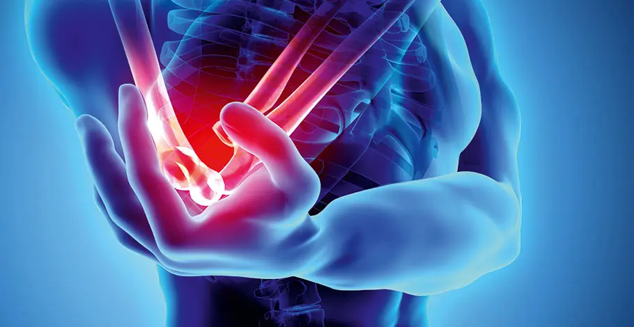
Artificial elbow joint
Compared to hip and knee wear on the elbow is rather rare, as the joint does not bear weight. Nevertheless, osteoarthritis can also occur here. It usually begins in the joint between the humerus and radius and then spreads to the other parts of the joint. Each year, orthopaedists orthopaedists and trauma surgeons in Germany implant around 400 elbow prostheses.
In the past, the elbow prosthesis was primarily used in the treatment of rheumatoid arthritis. Nowadays, this indication is less common due to improved drug therapies that slow down joint wear. Nevertheless, more implantations have been performed in recent years, especially to treat complex fractures of the lower part of the humerus or dislocation fractures in the area of the radial head.
In the case of elbow osteoarthritis, the Doctor not necessarily replace all joint parts. Depending on the clinical picture, previous injury, and stability of the collateral ligaments, he can also implant a partial endoprosthesis. In the early stage of osteoarthritis, it is sometimes sufficient to replace individual joint parts such as the radial head, the lower arm, or the surface of the radius. injury injury
Prosthesis models for the elbow
In the total endoprosthesis, the hinge joint between the upper arm and ulna is primarily replaced. Three types of elbow prosthesis models are generally available. An uncoupled prosthesis only replaces the surfaces of the worn-out joints. It can be used in young patients as long as the collateral ligament apparatus is stable (essential requirement!). A coupled prosthesis is used when the ligament apparatus is no longer sufficiently stable. Here, a partial axis or hinge ensures the stable guidance of the joint parts between the upper and lower arm. This type of prosthesis is very stable due to the axis guidance but loosens more quickly.
The most modern implant is the so-called partially coupled prosthesis, which guarantees improved stability through a "loose" joint connection in the form of a coupling mechanism but still allows a certain amount of play in the joint. This results in a longer lifespan (service life) and a very good functional outcome. Orthopedists today primarily implant partially coupled prostheses (for patients >65 years).
The procedure
The implantation is performed under general anesthesia in a supine or lateral position. Surgeons distinguish between two approaches: triceps-on or triceps-off. Triceps-on means that the doctor approaches the joint from behind without detaching the triceps muscle tendon. Triceps-off refers to the detachment of the triceps tendon from the bone. Experts today prefer the triceps-on procedure because the post-treatment and rehabilitation of patients can take place early and quickly. A triceps weakness or rupture of the triceps tendon can be prevented. The prosthesis is anchored with bone cement in the upper arm or ulna. Risks of joint replacement include infections, non-infectious prosthesis loosening, bone fractures, and nerve lesions.
Risks of a joint replacement surgery
The renowned medical journal Lancet described joint replacement in 2007 as the "operation of the 21st century." Nowadays, prosthetics typically last around 15 to 20 years, often longer. These numbers are steadily rising: Between 1993 and 2003 alone, the proportion of hip endoprostheses that still functioned well after 10 years increased from 92 to 95 percent. In addition to the durability of the prosthetic, the knowledge of potential complications is interesting for the long-term success evaluation of an artificial joint. Some of the most serious risks of joint replacement surgery include infections (early, late; superficial, deep), the formation of blood clots in a leg vein (thrombosis, possibly embolism), and so-called aseptic prosthetic loosening (without infection).
Crucial for the prognosis in individual cases is the earliest possible detection of a complication and its immediate, appropriate treatment. Infections require treatment with possibly specially selected antibiotics. For deep infections, partial or complete removal of the prosthetic may be necessary (see below). Thrombosis is detected through ultrasound examinations of the blood vessels and treated with medication that reduces the blood's Blood ability to clot. Annually, about 24,000 artificial hips and around 16,000 artificial knees are replaced in Germany. In about half of the cases, an infection is the underlying cause; beyond that, aseptic loosening (loosening without infection) and other mechanical causes are responsible for the prosthesis change.
The DGOOC (German Society for Orthopedics and Orthopedic Surgery) has certified operation centers (www.endocert.de) that ensure experienced surgeons oversee all procedures, all steps are documented, and staff is regularly trained. The case numbers of these centers are publicly accessible.
For a better outcome of artificial joint replacement
After an artificial joint replacement, pain-free Movement preferably without walking aids after six to eight weeks, or at the latest after three months. Appropriately dosed sports activities are not a problem in most cases (hiking, walking, golf, cycling, swimming, strength training, etc.). However, artificial joints are not sports joints and should be treated with care and caution. The better this is achieved, the higher the chances that an artificial joint will last for many years and, ideally, a lifetime. Modern high-tech implants and new surgical techniques help with this. Therefore, the behavior and lifestyle of the patient are also crucial for the success of the treatment. weight control, muscle building, coordination training (gait pattern), as well as the type and extent of everyday use are factors that lie entirely in the patient's hands.
prehabilitation
Preparation for joint replacement surgery is not solely in the hands of the treating physicians either. The patient themselves can do a lot before the procedure to make the surgeon's work easier and to achieve a better result. Weight Loss, Muscle strengthening, gait training, and the optimization of cardiovascular and metabolic values in consultation with the General practitioner ensure quicker surgeries, shorter anesthesia time, fewer complications, a shorter and more successful aftercare, as well as faster recovery.
Rehabilitation
An intensive rehabilitation lasting several weeks begins directly after the hospital stay (usually one week after surgery). It is intended to ensure a continuous healing process and a goal-oriented development of the patient's motor, cognitive, and psychological skills. Here, Pain is treated, wound healing is controlled, muscles are stabilized, coordination is trained, and self-confidence is rebuilt. The health condition, home care, and possible complications determine whether a patient undergoes rehabilitation in an inpatient (stay in the rehabilitation clinic with full board and therapeutic applications) or outpatient form (living at home, treatments in one or more therapeutic practices). The application for rehabilitation is handled by the hospital's social service in coordination with the surgeon and patient, with costs covered by the health insurance.
The follow-up care for a joint replacement surgery
Patients should not fully load an elbow prosthesis even after complete healing. They should lift a maximum of five kilograms; repetitive movements such as crafting or cleaning, as well as striking movements, should be avoided. For optimal results, an outpatient or inpatient rehabilitation usually follows the hospital stay, where patients mainly train the muscles surrounding the elbow joint, as well as the coordination and mobility of the joint. The rehabilitation phase generally lasts three to six months.
The elbow joint
The elbow joint connects the upper arm and forearm and functions as a pivot and hinge joint. The three joint components - humerus, ulna, and radius - allow a combined range of motion of the forearm and hand, as we need in everyday life and for various sports activities. Bony structures, ligaments, and muscles ensure the joint is stably guided. Due to this anatomical complexity, the elbow joint is particularly susceptible to overload damage, but especially to traumatic injuries. An elbow dislocation, for example, is the second most common joint dislocation (occurrence: 3-9 cases / 100,000 people) of adults after the shoulder, and insufficient treatment can lead to joint destruction and premature joint wear. Young adults are particularly susceptible to changes in the elbow joint due to sports-related overuse.
Arthritis and arthritis
Doctors differentiate between bony diseases and soft tissue injuries at the elbow. Bony diseases include elbow osteoarthritis and arthritis (joint inflammation). Elbow osteoarthritis usually affects people who have had a bone fracture, joint dislocation, or another bone disease. As a result of joint wear, cartilage damage, free joint bodies, bony growths, and deformities occur. In arthritis and rheumatic diseases, chronic inflammation destroys the joint.
Soft tissue injuries include overstretched and torn ligaments after joint dislocation, overloaded tendons, and muscular injuries. They can be accompanied by pain and restriction of movement up to elbow stiffness.
Experts for this Treatment Method

- Innovative Orthopedics & Sports Orthopedics
Dr. med. Martin Volz & Kollegen
Sportklinik Ravensburg
- Innovative Orthopedics & Sports Orthopedics
Prof. Dr. Hans-Georg Simank
Zentrum für Orthopädie und Neurochirurgie
- Innovative Orthopedics & Sports Orthopedics
Prof. Dr. med. Sebastian Siebenlist
Sektion Sportorthopädie TU München, Klinikum rechts der Isar
- Innovative Orthopedics & Sports Orthopedics
PD Dr. Wolfgang Pötzl
Vulpius Klinik Bad Rappenau
- Innovative Orthopedics & Sports Orthopedics
Prof. Dr. med. Christoph Lohmann
Orthopädische Universitätsklinik MagdeburgAll Experts in this Department
Show All- Innovative Orthopedics & Sports Orthopedics
Niklas Haberstroh
MW - Praxis für Orthopädie & Sportmedizin
- Innovative Orthopedics & Sports Orthopedics
PD Dr. med. Daniel P. Berthold
OrthoCenter München
- Innovative Orthopedics & Sports Orthopedics
Dr. med. Florian Pfalzer
Sportpraxis Stuttgart / Health & Aesthetic Stuttgart
- Innovative Orthopedics & Sports Orthopedics
Dr. med. Ivo Breitenbacher
MVZ für Orthopädie Flugfeld
- Innovative Orthopedics & Sports Orthopedics
Dr. med. Yvonne Ebel
Orthopädie Wernau
- Innovative Orthopedics & Sports Orthopedics