© Magnific
September 27, 2026
Christine Bürg & Marianne Waldenfels
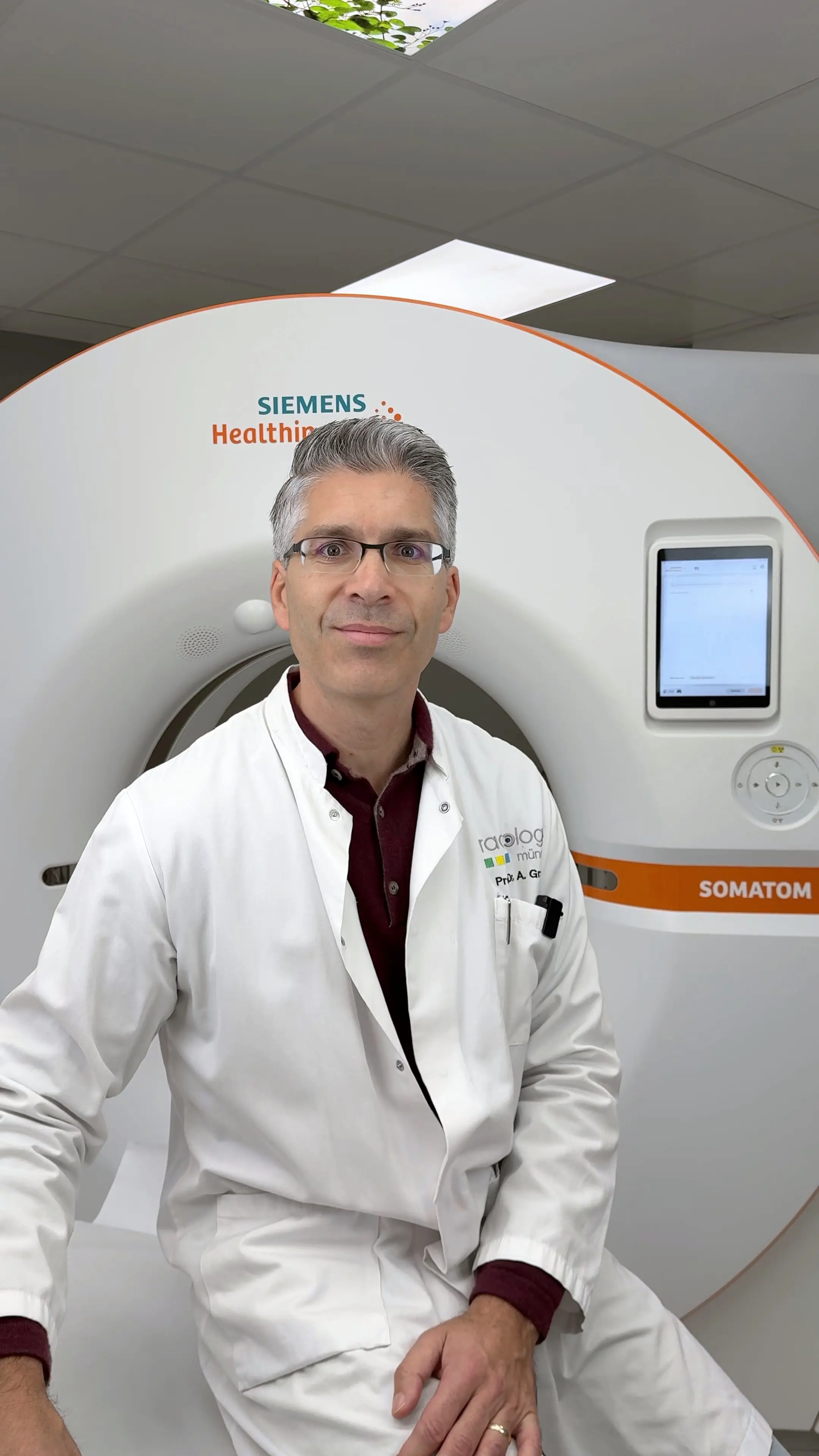
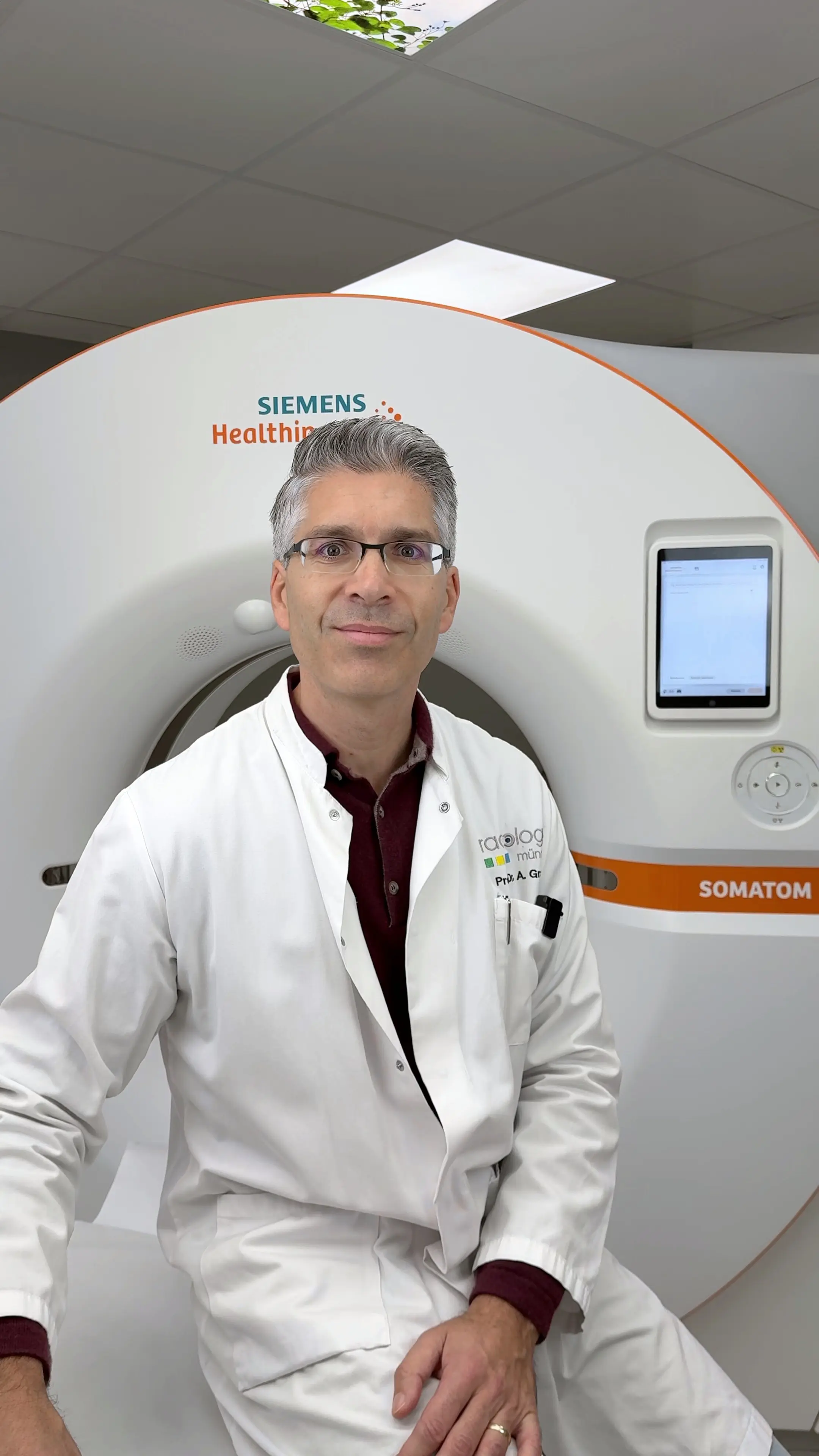
Finding cancer early can make a crucial difference – but the benefits of screening vary widely. Radiologist Prof. Dr. Anno Graser explains which methods can save lives and how CT, MRI and AI are changing cancer detection.

An interview with
Prof. Dr. Anno Graser
When it comes to early cancer detection, the logic seems straightforward at first: the earlier a tumor is found, the better. But it's not quite that simple. Some tumors might never have become dangerous, while others grow so fast that even an early diagnosis does not automatically confer an advantage.
Prof. Dr. Anno Graser, specialist in radiology and partner at Radiologie München, explains in this interview what CT and MRI can achieve today, when early detection saves lives – and what role AI might play in the future.
How early can cancer be detected today with modern imaging?
We can now detect some tumors in the millimeter range – often long before they cause any symptoms. That said, making a blanket statement like "cancer is visible five years in advance" would be irresponsible. It depends on the organ, the imaging method, and above all on the biology of the tumor.
It's not just about size. A small lung nodule, for example, stands out clearly against the air-filled lung tissue. A similarly small tumor in another organ, however, can look very much like healthy tissue. MRI additionally provides information about tissue structure, water molecule mobility, and blood flow, which helps us with classification.
An important distinction: seeing a change does not yet mean identifying it with certainty as cancer. And a small tumor is not automatically biologically early-stage. Our goal is to detect a dangerous disease at a stage where we can still decisively influence its course.
For which types of cancer do we truly know that early detection can save lives?
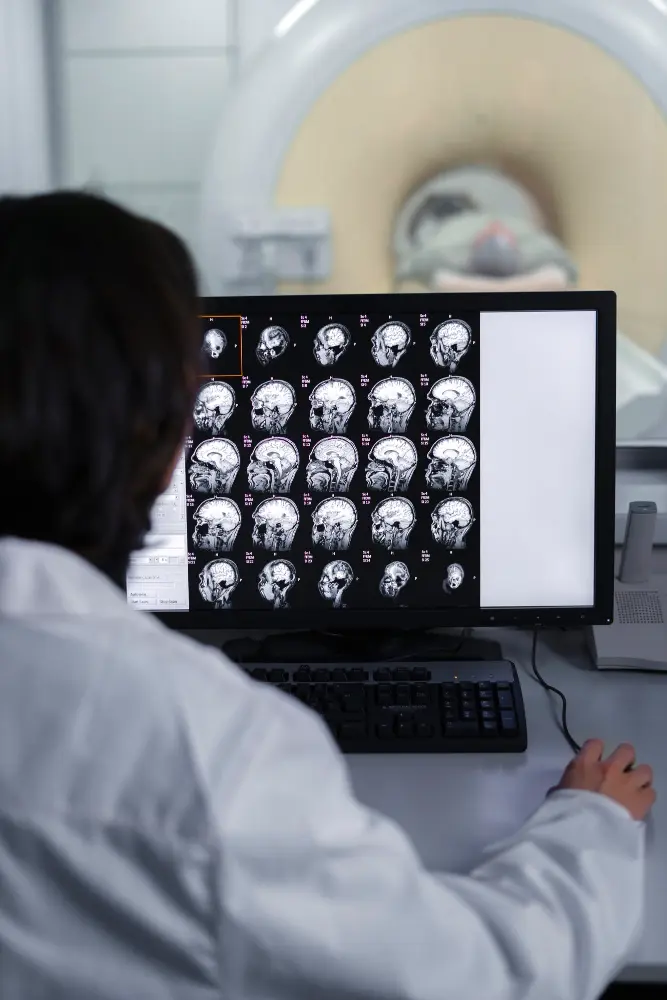
The most robust randomized evidence for radiological screening methods comes from mammography for breast cancer detection and low-dose CT of the lungs for people at elevated risk.
For lung cancer, large studies have shown that structured CT screening can reduce lung cancer mortality by approximately 20 to 24 percent in relative terms. This finding applies to the risk groups studied, not automatically to the general population. Since April 2026, via low-dose CT has been available for eligible statutory health insurance members. Requirements include being between 50 and 75 years of age and meeting certain criteria regarding duration of smoking and tobacco consumption.
With prostate cancer, a careful distinction must be made: for PSA-based early detection, there is long-term evidence of reduced prostate cancer mortality. MRI today improves the selection of candidates for biopsy and helps avoid unnecessary diagnoses. However, the additional mortality benefit of MRI itself has not yet been proven.
More early diagnoses are therefore initially a diagnostic success. Whether this also translates into fewer deaths must be investigated separately.
Where does imaging reach its limits?
Early detection is more difficult, for example, with pancreatic cancer and aggressive ovarian or fallopian tube carcinomas. Early changes can be extremely hard to detect in these cases, while the disease is already advancing biologically. Such tumors also frequently grow too fast for early detection to offer a real survival advantage.
CT and MRI also have limitations when it comes to superficial mucosal changes. For precancerous lesions and small tumors in the stomach, intestine, and urinary bladder, other methods – particularly endoscopy – are decisive. There is therefore no single imaging examination that reliably detects all types of cancer at an early stage.
Can you see tumors that would not have caused symptoms for years?
Yes. That is, on one hand, the opportunity of early detection, and on the other, its central challenge. We can detect tumors that would not cause symptoms until years later. In some cases, symptoms might not have developed at all within the patient's remaining lifetime.
This applies, for example, to certain slow-growing prostate carcinomas or individual very slow-growing lung tumors. The key point is: such a finding is not automatically a misdiagnosis. It may genuinely be cancer whose discovery still offers the person no health benefit. We call this overdiagnosis.
This is why "we've found something" must not automatically become "we must treat immediately." For suitable tumors, structured active surveillance can be the better strategy. The challenge lies in reliably distinguishing these cases from those where we cannot afford to lose time.
Can imaging determine which breast or prostate tumor requires treatment?
It can help us considerably with this distinction, but it cannot make the decision on its own. Imaging shows not only the size of a tumor but also its structure, blood supply, spread, and changes over time. From this, we can gain indications of its biological aggressiveness – though not a reliable individual prognosis for the future.
For the prostate, this progress is particularly well documented. With MRI, we can focus diagnostics more strongly on clinically relevant tumors and reduce unnecessary biopsies as well as the detection of less threatening tumors. However, a normal MRI does not completely rule out a relevant tumor.
For breast cancer, treatment decisions generally also require tissue examination and information about tumor biology. Overall, the decision to treat or actively monitor is made based on the interplay of imaging, histology, clinical risk, life expectancy, and the patient's wishes.
Why aren't far more people routinely examined by CT as a precaution?
Because the benefit of an examination depends not only on how good the equipment is, but also on how likely the condition being sought actually is in the people being examined.
In long-term smokers, we find lung cancer requiring treatment comparatively often. In people with very low risk, on the other hand, we find many harmless nodules and only a few dangerous tumors. This can lead to follow-up examinations, anxiety, and occasionally invasive procedures. Add to this overdiagnoses and a small but, with repeated CT examinations, non-negligible radiation exposure.
The conclusion is therefore not to examine as little as possible. We should better reach people with relevant risk and continue to improve selection. Good early detection consists of risk assessment, quality-assured imaging, and clear protocols for abnormal findings. A single CT appointment alone is not a screening program.
Can AI detect changes that a radiologist would not yet identify as cancer?
Yes, this is fundamentally possible, and for certain applications there is already convincing research data. AI can evaluate very subtle combinations of tissue structure, density, and spatial patterns that are difficult for the human eye to perceive.
However, we must distinguish between two things: Is the AI detecting an existing tumor that has been overlooked so far? Or is it detecting features that merely indicate an elevated risk of a future cancer? Medically, that is a significant difference.
A high risk score does not yet mean that cancer is already present. It would be particularly interesting to use this as a basis for individually adjusting examination intervals or for more targeted re-evaluation of existing scans. Before this becomes widespread practice, however, we must demonstrate that the results are reliable across other institutions and that the decisions derived from them genuinely help patients.
If you could make one decisive improvement to cancer early detection: where would you start?
I would choose low-dose CT for lung cancer early detection. Here we already have robust evidence that we can prevent deaths in people at elevated risk.
My most important improvement, however, would not simply be an even higher resolution. I would personalize the examination more consistently: who has a sufficiently high risk? Who needs which examination interval? And for which small lung nodule is a follow-up sufficient, rather than immediately initiating an invasive workup?
AI could support us with this risk assessment and with evaluating growth. Equally important are easy access for eligible individuals, reliable follow-up care, and smoking cessation support.
If I could improve just one thing, it would be the overall accuracy of the program: finding dangerous tumors in time while burdening as few people as possible with unnecessary workups.
The future of cancer early detection does not lie in finding more and more in every person. It lies in detecting dangerous diseases in time – and at the same time knowing as reliably as possible which changes we can monitor without needing to treat immediately.

"Feel good, live better" This podcast episode with Prof. Dr. Helen Abel is all about hands and how to keep them mobile – even with arthritis
Christine Bürg

An interview with

How often should beginners run, when should they increase their training, and how important are rest days? Physiotherapist Nils Stützer explains how to build a healthy running routine, what role running technique plays, and how to avoid overuse injuries.
Christine Bürg & Marianne Waldenfels

With
Nils Stützer

Finding cancer early can make a crucial difference – but the benefits of screening vary widely. Radiologist Prof. Dr. Anno Graser explains which methods can save lives and how CT, MRI and AI are changing cancer detection.
Christine Bürg & Marianne Waldenfels

An interview with
Prof. Dr. Anno Graser