Other Treatment Methods
- Dental anxiety / pain-free dental treatment
- Dental prosthesis
- Digital dentistry
- Gum treatment
- Orthodontics
- Pediatric dentist
- Prophylaxis / Professional Teeth Cleaning (PZR)
- Root canal treatment / Endodontics
- Sports dentistry
- Teeth grinding and CMD (Craniomandibular Dysfunction)
- Teeth whitening / Bleaching
- Veneers (dental shells)
Digital dentistry
The Digitization of our lives continues to progress steadily and in dentistry, this development also occupies a solid place. However, not always with satisfactory results! The digital dentistry is now an integral part of many treatment steps and indispensable in dental laboratories and dental practices. The rapidly changing possibilities must always be critically examined and tested, because precision and effort are not always in the necessary balance - especially when it comes to high-quality restorations.
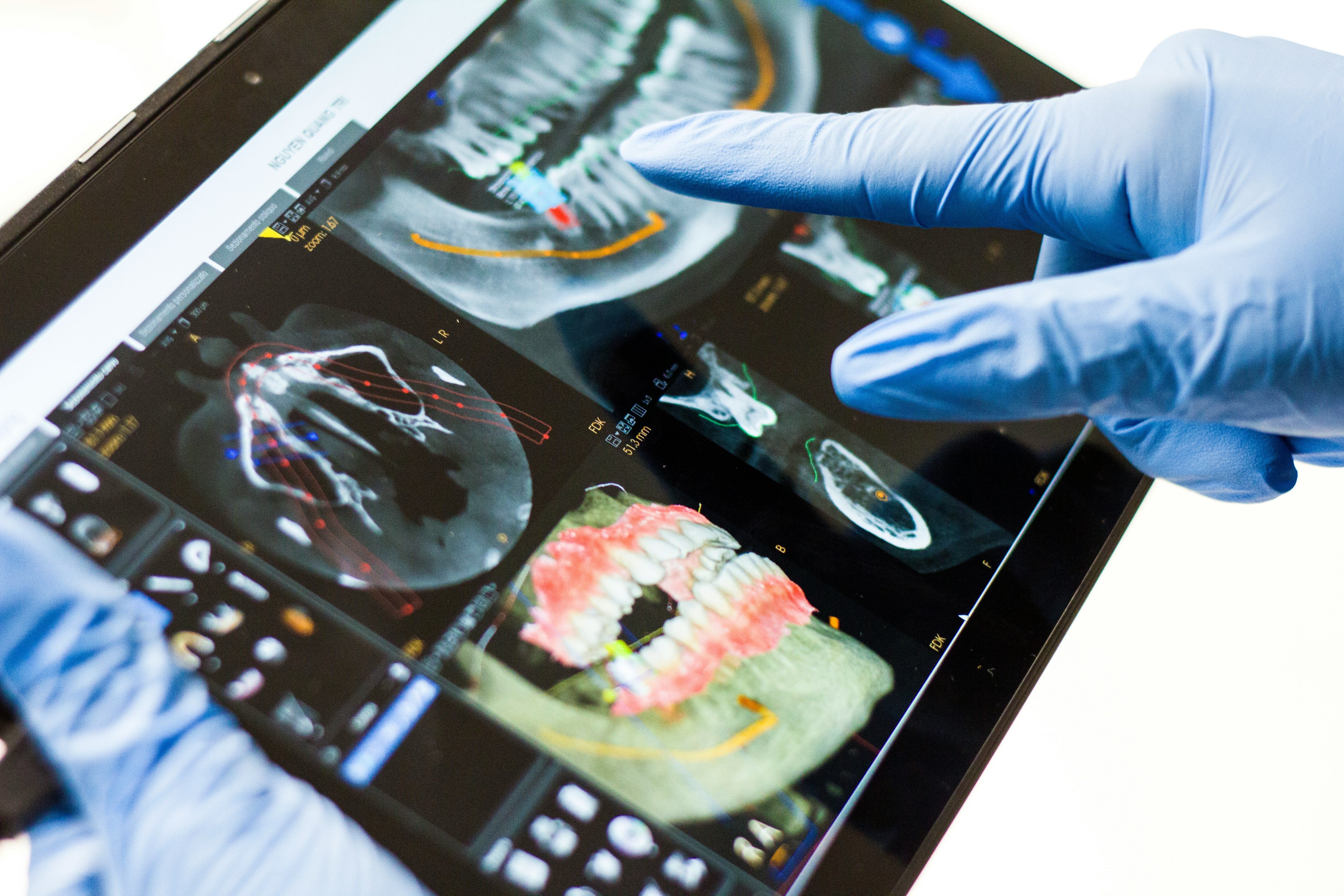
Low-radiation CBCT radiography (Cone Beam Computed Tomography)
Modern CBCT radiography enables a three-dimensional representation of the teeth and the jawbone and also provides insights into bone density and quality. In implantology the anatomical boundary structures - nerve pathways, nasal cavities, or maxillary sinuses - must be established in a three-dimensional representation in order to conduct an accurate analysis of the implant size and a possible bone augmentation before the procedure can be performed.
The teeth can also be represented in detail in three dimensions, which is absolutely necessary, for example, for root canal treatment. Because only the knowledge of the number and course of the tooth roots, as well as the detection of possible nerve branches, ultimately determines the success or failure of the treatment. In dental surgery too, DVT images offer an enormous advantage, especially for the course of surgical interventions, such as the removal of a wisdom tooth or an apicoectomy. If, for example, this is in the area of a molar tooth, the roots of which are in close proximity to sensitive nerve structures, the operation can be performed with significantly fewer problems if the three-dimensionality of the individual anatomy has already been analyzed in advance and can now be taken into account.
It is similar with the removal of an impacted tooth or the orthodontic necessary exposure of a so-called impacted canine tooth. Thanks to 3-D technology, the surgeon not only receives clear information about the surgical approach, accidental tongue-side perforations during implant drilling can also be avoided. This can happen if the bone structure in the rear part of the lower jaw is greatly reduced and this anomaly only becomes visible through digital volume tomography. It is clear that the surgical risk can be significantly reduced in all the aforementioned cases by including the DVT.
The use of DVT X-ray technology is very useful for many areas. In addition, in contrast to computed tomography (CT), it is significantly less radiation-intensive and also more cost-effective. In addition, digital processing allows for quick transmission and communication with all parties involved – dentists, dental technicians, patients, or general practitioners.
Digital evaluation and planning in implantology
The data obtained from the DVT can be transferred to special planning software. As a result, the available bone offer and the length and width of the implant are optimally adapted. Digitally milled implant templates are positioned and specified so precisely that minimally invasive implantation can take place in the correct position and drilling depth. As a result, the surgical intervention is gentle, fast, and with fewer healing complications.
Aesthetic analyses digital
Today, dentistry is able to offer a complete digital workflow. This means that all intraoral and extraoral information of the patient (i.e., everything inside and outside the oral cavity) is recorded digitally and made available for evaluation and therapy.
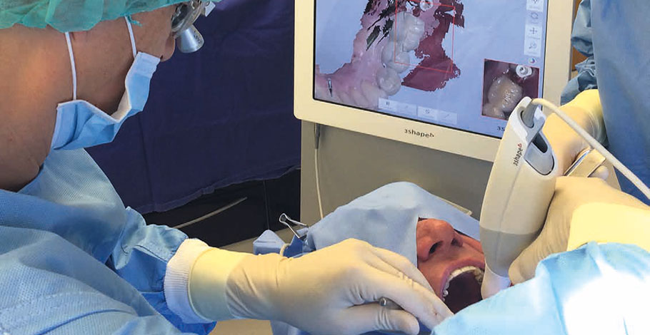
The face is first scanned three-dimensionally with a face scanner. This data can then be matched and overlaid with the DVT data. Additionally, an intraoral scan is performed to represent the upper and lower jaws. An electronic joint path recording then transmits movements digitally into the face and intraoral scan, so a real movable recording of the chewing act and the structures to be considered is created. In addition, special analysis software products can display an individual smile, which can be changed and developed together with the patient. This so-called Digital Smile Design (DSD) already gives a pretty good impression in advance of what the result could look like.
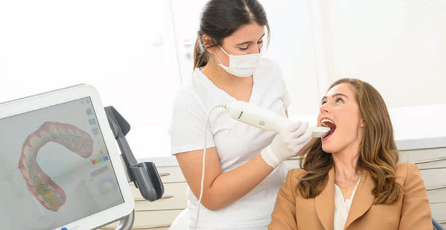
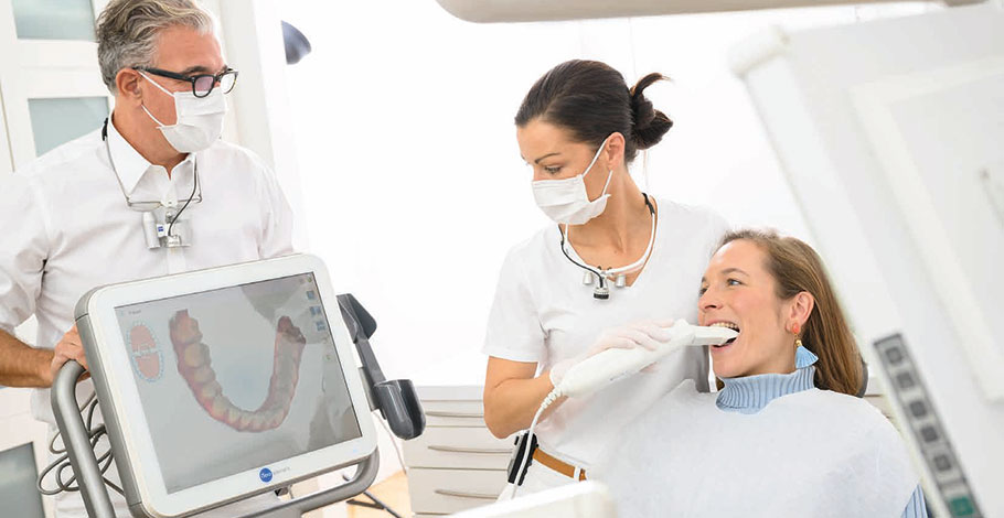
Intraoral scanners replace dental impressions
The fastest developing digital technology in dentistry is the intraoral scan, with which all structures in the oral cavity and teeth can be digitally recorded. For example, misalignments, overloads, changes and reductions in enamel and dentin or tissue loss can be accurately diagnosed. Prepared teeth or the position of implants can also be scanned and transmitted directly to the laboratory. The laboratory then manufactures the new crowns or bridges, inlays or partial crowns only using the digital data in the digital workflow.
All these representations and assessments therefore allow early intervention and thus secure the hard and soft tissue in their function in the mouth permanently and long-term. This technique has more difficulty when it comes to larger surfaces and several restorations. Here, the dimensional accuracy and precision of the analog analysis are unfortunately not yet equivalent. In addition, the costs for the acquisition and production of digital scanners and devices are not to be underestimated.
Progress control and documentation
A special advantage of digital dentistry is the good documentation and the possibility to quickly capture changes by overlapping the jaws. For example, after surgical or orthodontic changes, regular scans can help to quickly notice changes and intervene accordingly to correct them.
Other Treatment Methods in this Department
Experts for this Treatment Method
- Aesthetics & Function in Dentistry
Dr. Dr. Ákos Fehér
Fehér Dental Team, Smile Lounge
- Aesthetics & Function in Dentistry
Dr. med. dent. Malte Schönrock M.Sc., M.Sc.
Dent Aesthetic
- Aesthetics & Function in Dentistry
Jan Kurtz-Hoffmann
Zahnärzte im Roßbachpalais
- Aesthetics & Function in Dentistry
Dr. med. dent. Tore Thomsen
Zahnarztpraxis Dres. Thomsen & Kollegen
- Aesthetics & Function in Dentistry
Dr. med. dent. Oliver Brendel
Dinkelacker & Brendel
- Aesthetics & Function in Dentistry
Dr. med. dent. Nico Lindemann
Zahnärzte im RoßbachpalaisAll Experts in this Department
Show All- Aesthetics & Function in Dentistry
Dr. Dr. Ákos Fehér
Fehér Dental Team, Smile Lounge- Aesthetics & Function in Dentistry
Dr. med. dent. Malte Schönrock M.Sc., M.Sc.
Dent Aesthetic- Aesthetics & Function in Dentistry
Jan Kurtz-Hoffmann
Zahnärzte im Roßbachpalais- Aesthetics & Function in Dentistry
Dr. med. dent. Tore Thomsen
Zahnarztpraxis Dres. Thomsen & Kollegen- Aesthetics & Function in Dentistry
Dr. med. dent. Oliver Brendel
Dinkelacker & Brendel
- Aesthetics & Function in Dentistry