Other Treatment Methods
© PMC
Functional Urology
Functional urology is a subfield of urology that deals with disorders of the function of the lower urinary tract - i.e., problems with storing and emptying urine. Unlike tumors, inflammations or malformations, the focus here is not primarily on anatomical changes, but on functional disorders, often with complex causes.
Incontinence
Incontinence means that a person cannot or cannot fully control their urine (urinary incontinence). It is therefore an unintentional loss of urine. Urinary incontinence can manifest in different ways: In stress incontinence, also known as stress incontinence, urine loss occurs when there is pressure on the bladder. For example, when coughing, sneezing or lifting heavy objects. Urgency incontinence, on the other hand, is a sudden, strong urge to urinate with immediate urine loss. Mixed incontinence is a combination of both forms mentioned above, while overflow incontinence occurs when the bladder overflows when it is too full (e.g., with an enlarged prostate). If nerve control is disturbed, for example in a spinal cord injury, it is called reflex incontinence, i.e., involuntary emptying of the bladder.
Diagnosis
During a doctor’s visit, a physical examination is carried out in addition to taking the medical history, and urodynamics if necessary. Urodynamics is a medical examination that checks the function of the bladder and urethra – in particular, how well they can store and empty urine. This examination includes measuring bladder pressure, urine flow, and the urethral pressure profile. To do this, a thin probe is inserted into the bladder via the urethra. The bladder is then filled with liquid, while pressure conditions and the patient's reactions are documented simultaneously. The examination is somewhat uncomfortable, but generally not painful. It usually lasts 30–60 minutes.
Treatment methods
Conservative treatment methods include pelvic floor training to strengthen the muscles, as well as bladder training, i.e., regular toilet trips to train the bladder. Dietary adjustments can also be helpful, such as fluid management. In addition, medications can help, as well as electrical stimulation, biofeedback, and magnetic stimulation for muscle strengthening. Botox injections into the bladder can help with urgency incontinence; for stress incontinence in women, a so-called TVT or TOT band is often used. In this minimally invasive procedure, a plastic band is placed under the urethra to support it. In men, the so-called sling implantation is used. This procedure aims to heal stress urinary incontinence by compressing the urethra or realigning it with special material at its position to the bladder neck. Both techniques aim to prevent unwanted urine loss. The sling also supports the pelvic floor muscles, thus increasing resistance to the pressure of a full bladder.
Bladder emptying disorder
A bladder emptying disorder occurs when the bladder cannot be fully or only with difficulty emptied. This can lead to residual urine (i.e., urine remaining in the bladder), constant urge to urinate, dribbling of urine, or in extreme cases, urinary retention, where the bladder can no longer be emptied at all.
Causes and Symptoms
Causes of voiding disorders include impaired nerve supply to the bladder (for example in multiple sclerosis or Parkinson's disease) as well as mechanical disorders such as an enlarged prostate, urethral stricture, bladder stones or prolapse (in women). Medications such as antidepressants or sedatives can also inhibit bladder function, as well as a “relearned inability to urinate” such as after surgery. Which symptoms occur? In most cases, patients report a weak urine stream, frequent urination in small amounts, dribbling or feeling unable to completely empty themselves.
Diagnosis and Treatment
Here, the treating physician can choose among several procedures, such as residual urine measurement via ultrasound, urodynamics, cystoscopy, and neurological examinations. Patients are also often advised to keep a bladder diary. In conservative therapy, bladder training (regular emptying without urge) and double voiding (attempting to void again a few minutes later) can be recommended. Especially with functional disorders, pelvic floor training and/or physiotherapy help. Medications such as alpha-blockers relax the bladder neck and prostate and support bladder contraction. In the very proven method of ISC (intermittent self-catheterization), the patient empties the bladder regularly with a single-use catheter; an indwelling catheter is only recommended in special situations.
Neurogenic Bladder Dysfunction
Neurogenic bladder dysfunction means that the bladder no longer works properly due to nerve damage. What exactly does this mean? Bladder filling and emptying are controlled by the nervous system, but when this control is disrupted, incontinence, voiding disorders, or both can occur. A healthy bladder control always works on the same principle: the bladder fills, expands, nerve signals are sent to the brain, a conscious urge to urinate arises. When urinating, the brain allows emptying, the bladder muscle contracts, the sphincter muscle relaxes and urine flows out.
Neurogenic bladder disorder is differentiated into three forms: In the reflex bladder, also called spastic bladder, the bladder contracts uncontrollably, resulting in urge incontinence. The areflexive bladder (flaccid bladder) no longer contracts properly; the consequences are urinary retention, overflow incontinence and residual urine. In dyssynergy between the bladder and sphincter muscle, the bladder contracts but the sphincter muscle no longer opens. This leads to impaired emptying with high bladder pressure.
Causes and Symptoms
There are various causes for impaired bladder function, such as multiple sclerosis, Parkinson's, a stroke, diabetic neuropathy or slipped discs. Typical effects include delayed urge to urinate, coordination disorders, lack of urge to urinate or pressure on the spiral nerves; but also urinary tract infections or back pain when the kidneys are congested.
Diagnosis and Treatment
The specialist has various test procedures available for diagnosis. In addition to a thorough medical history, a residual urine measurement can be performed via ultrasound. Urodynamics is also used, as well as a cystoscopy, also known as a cystoscopy. Complementary neurological tests and imaging procedures such as an MRI are used. Depending on the findings, medication may be used for a spastic bladder or to relax the bladder neck. An overactive bladder is treated with Botox, and SIC (intermittent self-catheterization) is also particularly effective here. Other proven measures include bladder training, fluid management, physiotherapy, toilet training and regular check-ups to protect kidney function.
Diseases of the urinary tract
Diseases of the urinary tract affect the organs responsible for the production, storage, and elimination of urine. These include the kidneys, ureter, bladder, and urethra.
Urinary tract infection
A urinary tract infection (UTI) is an inflammation caused by microorganisms and affects one or more parts of the urinary tract. A UTI manifests itself either as cystitis (inflammation of the bladder), urethritis (inflammation of the urethra), or pyelonephritis (inflammation of the renal pelvis).
Causes and Symptoms
The main cause of a UTI is in most cases the penetration of intestinal bacteria, less commonly viruses or fungi, which migrate upwards towards the bladder through the urethra. Which risk factors favor a UTI? In women, for example, a short urethra that is close to the anus, in men, an enlarged prostate. Classic "honeymoon cystitis" (through sexual activity), hypothermia, a weakened immune system, or diabetes can also be triggers.
Depending on where the urinary tract infection is located, the symptoms vary. If the lower urinary tract (cystitis, urethritis) is affected, burning during urination, frequent urge to urinate, pain in the lower abdomen or cloudy urine occur. If the upper urinary tract (pyelonephritis) is affected, there is a general feeling of illness, as well as pain in the side, fever, chills, or nausea.
In addition to classical cystitis, i.e., a bladder infection, there is also interstitial cystitis, a chronic bladder pain syndrome. This non-bacterial inflammation of the bladder is of unclear cause and is associated with pain. The symptoms resemble cystitis - chronic urge to urinate, bladder pain, and small amounts of urine. Since there are no germs in the urine, further diagnostics by the urologist are necessary.
Diagnosis and treatment
In addition to medical history and physical examination, a urine test is performed. If pyelonephritis is suspected, a blood test for inflammatory markers is also conducted. In case of suspected drainage problems or urinary stones, an ultrasound is used. Depending on the severity of the inflammation, a multi-day antibiotic therapy is prescribed. It is also important to drink plenty of fluids.
Overactive bladder
Overactive bladder, also known as OAB (Overactive Bladder), is a functional bladder disorder characterized by excessive activity of the bladder muscle (detrusor) without any identifiable organic or infectious cause.
Causes and symptoms
The exact cause is not always clear, but there are some possible triggers, such as nerve overactivity in the bladder receptors, chronic stress, estrogen deficiency (keyword menopause), overweight, or a past bladder infection. However, the symptoms are usually clearly recognizable: sudden, strong urge to urinate and noticeably frequent urination during the day as well as increased nighttime urination. In some cases, there is also what is known as urge incontinence, i.e., urinary leakage when feeling the urge to urinate.
Diagnosis and treatment
The attending physician will first take a medical history and examine the urine to rule out a urinary tract infection. An ultrasound can evaluate residual urine, bladder wall, and kidneys. If necessary, urodynamics may also be initiated. A symptom diary in which the patient records drinking amounts, toilet visits, and urine output can also be helpful. With regard to therapy, there are several options: Non-drug measures include pelvic floor training, bladder training (deliberately delaying bladder emptying to train stretching capacity), and reducing or avoiding irritating substances such as coffee, alcohol, nicotine, and spicy foods. In terms of pharmacological therapy, anticholinergics help calm an overactive bladder muscle, beta-3 adrenergic receptor agonists promote bladder relaxation, and Botox injections into the bladder muscle paralyze overactivity.
Bladder stones
Bladder stones are solid crystal formations in the bladder that form from components of urine. They can occur singly or in multiples, be as small as grains of sand, or as large as golf balls. They form either primarily in the bladder or as a result of descending kidney or ureter stones.
Causes and symptoms
Since bladder stones occur more frequently in men, the causes are usually an enlarged prostate, a neurogenic bladder (a bladder emptying disorder), a narrowing of the urethra, or foreign objects in the urethra such as an indwelling catheter. Some bladder stones remain asymptomatic for a long time, while others cause symptoms such as pain during urination, frequent urge to urinate, blood in the urine, or 'gritty' urine (a stone in the urine).
Diagnosis and treatment
First, the medical history and physical examination are performed. The urine is examined for blood, crystals, and signs of infection. Imaging techniques such as ultrasound, X-ray, CT, or cystoscopy allow precise localization of the stones. Therapeutically, small, asymptomatic stones are initially only monitored. Increased drinking often leads to spontaneous passage. Larger or symptomatic stones can be removed endoscopically, minimally invasive through the urethra, or shattered with laser or ultrasound. Shock wave lithotripsy can also be used. Very large stones may need to be surgically removed. Accompanying infections are treated with antibiotics.
Urethritis
The urethra is a relatively small but important organ of the urinary tract and can be affected by various diseases. Urethritis, inflammation of the urethra, is most commonly caused by infections.
Causes and symptoms
The inflammation is usually caused by various bacteria, such as Chlamydia. Typical symptoms include burning during urination, frequent urge to urinate, itching, or a whitish discharge from the urethra.
Diagnosis and treatment
The doctor will conduct a urine test (first stream), perform a urethral swab, and carry out targeted PCR tests for Chlamydia, gonococci, and other pathogens. Depending on the findings, the appropriate antibiotics will be prescribed.
Urethral stricture
In the case of a so-called urethral stricture, a narrowing of the urethra, it is usually due to a malformation or a scarred change in the tissue.
Causes and symptoms
In addition to a congenital malformation, injuries, such as those caused by a catheter, or inflammations can also be triggers. Typical symptoms include a weak urinary stream, dribbling, or the feeling of incomplete bladder emptying.
Diagnosis and treatment
The urologist will order a urine flow measurement in case of suspicion, but diagnoses can also be made using a urethrogram or an endoscopy. Treatment is carried out, for example, through catheter therapy or by careful dilation of the urethra.
Urethral injury
Trauma to the urethra can also occur due to traffic accidents, falls, external violence, or a catheter. Treatment must then be discussed individually with the attending physician.
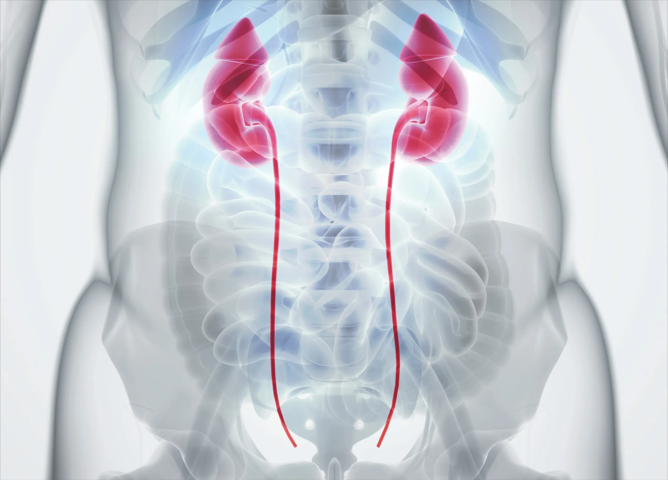
Ureteral diseases / Drainage disorders
The ureters are two thin muscle tubes that transport urine from the kidneys to the bladder. They can also be affected by functional disorders, usually in the form of drainage disorders. These are conditions in which urine flow between the kidney and the bladder is obstructed. Such a backup of urine can have serious consequences, such as hydronephrosis, infections, or long-term kidney damage.
Urinary retention
Urinary retention (hydronephrosis or urinary drainage disorder) refers to the backup of urine in the upper urinary tract, usually in the ureters and renal pelvis, because urine cannot flow freely from the kidney to the bladder.
Causes and symptoms
A distinction is made here between mechanical causes, such as ureteral stones, an enlarged prostate, scars, or tumors, and functional causes such as urine reflux. As with almost all urinary tract diseases, the symptoms manifest as flank pain, blood in the urine, or acute urinary retention.
Diagnosis and treatment
Urinalysis, ultrasound, and imaging procedures such as CT or MRI are used for clarification. The goal of treatment is to resolve acute urinary obstruction, for example, using a catheter that is placed directly into the kidney. Medication therapy accompanies the procedure to relieve pain and inflammation.
Ureteral stones (Ureterolithiasis)
Ureterolithiasis refers to a kidney stone descending into the ureter and causing urinary obstruction.
Symptoms
Acute colicky flank pain that typically occurs in waves is one of the classic symptoms. Blood in urine, nausea, and vomiting can also indicate the formation of ureteral stones.
Diagnosis and treatment
Clarity is usually provided by a urinalysis and an ultrasound. Treatment involves painkillers, medical stone dissolution, or ureteroscopy, where the stone is fragmented and removed.
Ureteral narrowing (Ureteral stricture)
When the ureter is narrowed, urine flow is obstructed, leading to urinary retention.
Causes and Symptoms
Scars or tumors are often the cause of a narrowing, less commonly inflammations. Typical indications of a ureteral stricture are recurrent urinary tract infections, urinary retention, and flank pain.
Treatment
A balloon catheter is inserted into the ureter and positioned at the narrowed area. There the balloon gently expands and widens the narrowed section. In some cases, the ureter may also be surgically reconstructed.
Kidney Stones
Kidney stones (nephrolithiasis) are solid crystalline deposits that form from components of urine in the kidneys. They can be as small as grains of sand or several centimeters large. Overall, kidney stones are common but well treatable – a swift diagnosis and an individually tailored therapy are crucial to alleviate symptoms and maintain kidney function in the long term.
Causes and Symptoms
The causes of kidney stones are diverse: insufficient fluid intake, unbalanced diet, metabolic disorders, genetic predisposition, lack of exercise, urinary tract infections, or anatomical peculiarities in the urinary tract can promote stone formation. While small stones often remain symptom-free and pass unnoticed with the urine, larger ones can obstruct urine flow or migrate into the ureter. This often leads to severe, colicky flank pain — the typical renal colic — often accompanied by nausea, vomiting, blood in the urine, and increased urge to urinate.
Diagnosis and Treatment
Diagnosis is usually made using ultrasound and computed tomography (CT), allowing for precise localization and size assessment of the stone. Additionally, urine is examined for blood, signs of inflammation, and crystals. Therapy is then based on the size, location, and composition of the stone, as well as the individual symptoms of the patient.
Small stones up to about 5-6 mm can often be passed naturally, supported by a high fluid intake, pain relievers, and possibly medications to relax the muscles of the ureter. The goal of this conservative therapy is the spontaneous passage of the stone. Larger or painful stones require further interventions. These include extracorporeal shock wave lithotripsy (ESWL), where the stone is shattered by sound waves, and ureteroscopy, where the stone is removed endoscopically via the urethra and bladder or fragmented with a laser. In the case of very large or complicated stones, percutaneous nephrolitholapaxy (PNL) may be necessary, meaning direct removal from the kidney via a small skin incision.
Kidney cysts
Kidney cysts are fluid-filled cavities that form in the renal tissue. They are among the most common changes in the kidneys and are usually benign. Generally, they occur singly or sporadically and cause no symptoms. These are referred to as simple kidney cysts, which are often discovered incidentally on ultrasound, for example during a check-up.
Causes and symptoms
The exact cause of kidney cysts is not fully understood, but they are more common in older age. In people over 50, about half have at least one cyst in the kidney. These cysts are surrounded by a thin wall, contain clear fluid, and are not related to tumors. Small cysts usually cause no or only mild symptoms. However, larger cysts (from about 5 cm) can exert pressure on surrounding tissue and cause a pulling sensation in the flank. Complications such as bleeding, infections, or ruptures of the cyst are rare.
Diagnosis and treatment
Diagnosis is usually done via ultrasound; for a more detailed assessment, a CT or MRI scan may additionally be performed. Treatment is based on the symptomatology. Asymptomatic cysts do not require therapy but should be monitored by ultrasound every 6 to 12 months. Symptomatic cysts can be punctured and drained; in some cases, a sclerosing agent is injected to shrink the cyst wall. In rare cases, such as recurrent symptoms or very large cysts, surgical (laparoscopic) removal may be appropriate.
Renal malformations
Renal malformations are congenital developmental disorders of the kidneys that occur before birth and can vary greatly in their manifestation. They are among the most common malformations of the urinary tract and can occur singly or bilaterally. Some of these changes remain unnoticed for a lifetime, while others lead to complaints or dysfunctions in childhood. The most common forms include the complete absence of one or both kidneys, a reduced-size kidney, or a pelvic kidney (where the kidney is located lower than usual).
Symptoms
The symptomatology is heavily dependent on the type and extent of the malformation. Many malformations remain asymptomatic and are only discovered by chance. Others can cause recurring urinary tract infections, urinary outflow obstructions, high blood pressure, or, in the worst case, impaired kidney function. Children with more complex malformations sometimes show early growth disturbances or bladder problems.
Diagnosis and treatment
The diagnosis is often made during prenatal ultrasound examination or in the newborn or early childhood stage. Imaging techniques such as ultrasound or MRI allow for an accurate assessment of the anatomical and functional conditions. Blood and urine tests provide additional clues about infections or loss of function. Depending on the type of malformation and the associated symptoms, the doctor determines the appropriate therapy. Mild forms often require no treatment but only regular follow-up examinations. Urinary outflow obstructions can be surgically corrected, for example, by a pyeloplasty in the case of flow restrictions. Double ureters or reflux may also require surgical correction. In severe bilateral malformations, long-term renal replacement therapy (dialysis, kidney transplantation) may be necessary.
Overall, many kidney malformations can be well controlled if detected early, some even heal completely or never require treatment. Crucial is targeted and individually tailored medical care.